
ABSTRACT
Introduction and objectives: Transcatheter implantation of balloon-expandable valves in non-aortic positions represents a growing off-label strategy for valve-in-valve and valve-in-ring procedures in high-risk patients. We assessed 4-year clinical outcomes and echocardiographic performance of the transcatheter heart valve Myval THV implanted in the mitral, tricuspid, and pulmonary positions.
Methods: This retrospective multicenter study included a total of 50 patients (mitral n = 33; tricuspid n = 13; pulmonary n = 4) treated across 11 centers, with clinical follow-up extending to 4 years and echocardiographic assessment of surviving patients.
Results: Technical successs was achieved in 96.9% of mitral cases (1 procedural death) and in 100% of tricuspid and pulmonary cases. At 1 year, death had occurred in 2/33 (6.1%) mitral patients and in none of the tricuspid or pulmonary patients. At 4 years, death had occurred in 4/33 (12.1%) mitral, 2/13 (15.3%) tricuspid, and 0/4 pulmonary patients; stroke occurred in 0/33, 1/13 (7.6%), and 0/4 patients, respectively. Mean transvalvular gradients at 4 years were 8.5 ± 3.3 mmHg (mitral), 3.8 ± 1.6 mmHg (tricuspid), and 12.7 ± 9.9 mmHg (pulmonary). No moderate or greater bioprosthetic regurgitation was observed at 4-year echocardiographic assessment, and no valve reinterventions were recorded during follow-up.
Conclusions: In this multicenter cohort, off-label use of the Myval THV in non-aortic positions demonstrated adequate midterm clinical outcomes and sustained echocardiographic performance up to 4 years. These preliminary findings support the feasibility of this transcatheter approach in selected patients but require further prospective validation.
Keywords: Myval. Transcatheter heart valve. Mitral valve-in-valve. Tricuspid valve-in-valve. Pulmonary valve-in-valve. Valve-in-ring. Long-term outcomes.
RESUMEN
Introducción y objetivos: El implante percutáneo de válvulas expandibles con balón en posiciones no aórticas representa una indicación fuera de ficha técnica cada vez más utilizada para procedimientos de válvula sobre válvula (valve-in-valve) y valvula sobre anillo (valve-in-ring) en pacientes de alto riesgo. Se evaluaron los resultados clínicos a 4 años y el seguimiento ecocardiográfico de la prótesis valvular cardiaca percutánea Myval THV implantada en las posiciones mitral, tricuspídea y pulmonar.
Métodos: Este estudio multicéntrico retrospectivo incluyó a 50 pacientes (mitral, n = 33; tricuspídea, n = 13; pulmonar, n = 4) tratados en 11 centros, con seguimiento clínico hasta 4 años y evaluación ecocardiográfica de los pacientes supervivientes.
Resultados: Se alcanzó éxito técnico en el 96,9% de los casos mitrales (1 muerte intraoperatoria) y en el 100% de los tricuspídeos y pulmonares. Al año, habían fallecido 2/33 (6,1%) pacientes del grupo mitral y ninguno de los pacientes de los grupos tricuspídeo ni pulmonar. A los 4 años, habían fallecido 4/33 (12,1%) pacientes del grupo mitral, 2/13 (15,3%) del tricuspídeo y 0/4 del pulmonar; se registraron ictus en 0/33, 1/13 (7,6%) y 0/4 pacientes, respectivamente. Los gradientes transvalvulares medios a los 4 años fueron de 8,5 ± 3,3 mmHg en la posición mitral, 3,8 ± 1,6 mmHg en la tricuspídea y 12,7 ± 9,9 mmHg en la pulmonar. No se observó insuficiencia bioprotésica de grado moderado o superior en la evaluación ecocardiográfica a 4 años ni se registraron reintervenciones valvulares durante el seguimiento.
Conclusiones: En esta cohorte multicéntrica, la indicación fuera de ficha técnica de la Myval THV en posiciones no aórticas mostró resultados clínicos adecuados a medio plazo y un rendimiento ecocardiogáfico mantenido hasta los 4 años. Estos hallazgos preliminares respaldan la viabilidad de este abordaje percutáneo en pacientes seleccionados y justifican su posterior validación en estudios prospectivos.
Palabras clave: Myval. Prótesis valvular cardiaca percutánea. Válvula sobre válvula mitral. Válvula sobre válvula tricuspídea. Válvula sobre válvula pulmonar. Válvula sobre anillo. Durabilidad.
BEV: balloon-expandable valve. THV: transcatheter heart valve. ViMAC: valve-in-mitral annular calcification. ViR: valve-in-ring. ViV: valve-in-valve.
INTRODUCTION
Balloon-expandable transcatheter heart valves (BEVs), originally developed for the treatment of severe aortic stenosis, have transformed the landscape of structural heart interventions and are now routinely used in patients across all surgical risk categories. While aortic valve replacement remains their primary indication, BEVs have seen increasing off-label application in non-aortic positions, particularly in the mitral, tricuspid, and pulmonary valves, where reoperative surgical risk is often prohibitive due to patient comorbidities, previous sternotomies, or hostile anatomy.1
In the mitral position, transcatheter valve-in-valve (ViV) procedures have become a viable alternative to redo surgery in patients with degenerated bioprosthetic valves. Early experiences have demonstrated acceptable procedural success and midterm survival, although challenges such as left ventricular outflow tract (LVOT) obstruction, device anchoring, and paravalvular leak (PVL) remain concerns.2 Valve-in-ring (ViR) procedures in mitral annuloplasty rings pose additional complexity due to variability in ring rigidity and geometry, which can impact valve expansion and sealing. Similarly, valve-in-mitral annular calcification (ViMAC) represents one of the most technically challenging scenarios due to irregular annular anatomy, risk of LVOT obstruction, and the absence of a defined anchoring structure.3
Tricuspid ViV and ViR procedures have emerged as bailout therapies for failed surgical tricuspid repair or replacement. The tricuspid annulus is typically large and elliptical, with lower pressures and complex three-dimensional geometry, making anchoring and positioning more challenging. Nonetheless, registries have shown favorable midterm valve-related outcomes with BEVs in this position.4
In the pulmonary position, transcatheter pulmonary valve replacement is an established treatment option for patients with right ventricular outflow tract conduit dysfunction, particularly in those with previous congenital heart surgery. BEVs have extensive experience in this setting and have demonstrated favorable clinical and hemodynamic outcomes, particularly in conduits and surgically placed bioprostheses.5-10
The Myval transcatheter heart valve (THV) (Meril Life Sciences, India) is a next-generation BEV featuring a hybrid honeycomb frame and dual internal/external polyethylene terephthalate skirts to reduce PVL. It uses bovine pericardial leaflets processed to mitigate calcification and has been CE-marked for use in the aortic position. Its design enables a broad range of diameters and fine sizing increments, which may offer advantages in non-aortic anatomies. Although Myval has been adopted in off-label settings across mitral, tricuspid, and pulmonary positions, data on long-term clinical and echocardiographic performance in these applications remain limited.1
This study aims to address this gap by presenting a retrospective, multicenter analysis of patients who underwent off-label Myval THV implantation in the mitral, tricuspid, and pulmonary positions, with 4 years of clinical and echocardiographic follow-up. Given the distinct anatomical and hemodynamic characteristics of these 3 settings, outcomes were assessed separately according to valve position.
METHODS
Study design and population
This retrospective, multicenter observational study included consecutive patients undergoing Myval THV off-label implantation in the mitral, tricuspid, or pulmonary positions from December 2017 through April 2020 at 11 centers in Europe and Asia. Patients were identified from the local structural heart intervention databases of the participant centers. Eligibility required Myval THV implantation in a non-aortic position for a degenerated surgical bioprosthesis, failed annuloplasty ring, or severe mitral annular calcification. Patients evaluated for these interventions who ultimately did not undergo Myval implantation were not systematically recorded; therefore, the number of potentially eligible patients, screening failures, and reasons for exclusion could not be retrospectively reconstructed. Reporting of this observational study followed the STROBE recommendations.11
Device description
The Myval THV device, Conformité-Européenne approved since April 2019, is a BE prosthesis with a nickel-cobalt alloy frame with hexagonal cells covered internally and externally in the ventricular end to minimize the potential for PVL. The second-generation Myval OCTACOR THV has 2 rows of identical octagonal cells that reduce the rate of foreshortening and a higher circumferential external skirt, designed to further decrease the risk of leakage. Both generations contain 3 leaflets composed of bovine pericardium tissue and are available in conventional (20 mm, 23 mm, 26 mm, and 29 mm), intermediate (21.5 mm, 24.5 mm, and 27.5 mm) and extra-large sizes (30.5 mm and 32 mm).12 Device size was selected based on three-dimensional multidetector computed tomography scans. All sizes are compatible with the 14-Fr Python introducer sheath (Meril Life Sciences, India). The Myval-1 study previously demonstrated the safety and efficacy profile of this valve in intermediate- to high-risk patients in the aortic position.12 The LANDMARK study showed that aortic Myval was noninferior to a contemporary prosthesis (SAPIEN [Edwards Lifesciences, United States], or Evolut [Medtronic, United States]).13
Procedural details
Procedural strategy was tailored to anatomy and valve position. Mitral procedures included transseptal access with preprocedural planning to mitigate LVOT obstruction risk, with laceration of the anterior mitral leaflet to prevent outflow obstruction (LAMPOON) performed where necessary. Access was predominantly transfemoral or transjugular. Balloon predilation was selectively used.
Outcomes and definitions
Primary endpoints included all-cause mortality, cardiac death, and stroke assessed at predefined 1- and 4-year follow-up time points. Secondary endpoints were echocardiographic valve-performance parameters (mean gradients, valve areas, and regurgitation severity) and valve-related adverse events, including thrombosis, endocarditis, migration, and valve reintervention. Technical successs for mitral interventions was defined according to the Mitral Valve Academic Research Consortium (MVARC) criteria and assessed at exit from the procedure room.14 For tricuspid and pulmonary interventions, analogous components of technical success were applied descriptively to provide consistency of procedural reporting, acknowledging that MVARC definitions were developed for mitral interventions and have not been specifically validated in these anatomical positions.
Follow-up
Clinical outcomes were assessed at predefined 1- and 4-year follow-up time points. Echocardiographic follow-up was performed according to local clinical practice. At each time point, echocardiographic analyses included patients alive with an available examination; patients who had died before the corresponding time point were not included in the echocardiographic denominator. Missing echocardiographic data were not imputed. Reasons for missing examinations were recorded when available but could not always be retrospectively established. Systematic computed tomography surveillance was not performed, and CT was obtained only when clinically indicated.
Statistical analysis
Categorical variables are expressed as absolute numbers and percentages and continuous variables as mean ± standard deviation (SD). Given the descriptive nature and limited sample size of the study, no formal comparisons between anatomical positions or procedural subgroups were performed. Clinical events were assessed at predefined 1- and 4-year time points and are reported as observed proportions with 95% exact binomial confidence intervals for major clinical outcomes. Exact dates of death and stroke were not available for all events; therefore, Kaplan–Meier or competing-risk analyses could not be reliably performed, and no event-time imputation was undertaken. Echocardiographic analyses were performed using available cases at each follow-up time point, and missing values were not imputed. All analyses were performed using R software (version 3.6.1, R Project for Statistical Computing, Austria).
Ethics
The study was conducted in full compliance with the principles outlined in the Declaration of Helsinki and was approved by the local Clinical Research Ethics Committee (CEIC) of the leading institution. Given the retrospective nature of the study, the requirement for specific informed consent for participation in the study was waived by the Ethics Committee. All procedures were performed in compliance with standard clinical practice, and informed consent for the procedure was obtained from all patients when applicable.
RESULTS
Baseline characteristics
A total of 50 patients underwent Myval THV implantation in a non-aortic position during the study period: 33 in the mitral position, 13 in the tricuspid position, and 4 in the pulmonary position. Patients undergoing mitral or tricuspid interventions had a substantially higher estimated surgical risk than those undergoing pulmonary intervention (Society of Thoracic Surgeons [STS] score 7.7 ± 5.7% vs 2.8 ± 0.9%, respectively; P < .001). Women represented 63.0% (29/46) of the combined mitral and tricuspid cohort compared with 50.0% (2/4) of the pulmonary cohort, although this difference was not statistically significant (P = .63). The mean age was 71.8 ± 12.3, 50.3 ± 16.2, and 48.1 ± 23.1 years for mitral, tricuspid and pulmonary interventions, respectively, reflecting the different clinical populations represented by the three anatomical groups. Main baseline characteristics of the study population are depicted in table 1. Within the mitral cohort, procedures included ViV, ViR, and ViMAC procedures; outcomes according to these specific mitral substrates are reported in table S1.
Table 1. Main clinical, electrocardiographic, and imaging characteristics at baseline of the study population
| Variables | Mitral (N = 33) | Tricuspid (N = 13) | Pulmonary (N = 4) |
|---|---|---|---|
| Patient characteristics | |||
| Age, years | 71.8 ± 12.3 | 50.3 ± 16.2 | 48.1 ± 23.1 |
| Female sex | 19 (57.6) | 10 (76.9) | 2 (50.0) |
| BSA, m2 | 1.73 ± 0.16 | 1.70 ± 0.23 | 1.67 ± 0.36 |
| BMI, kg/m2 | 25.0 ± 3.7 | 26.8 ± 3.5 | 23.7 ± 2.3 |
| Diabetes mellitus | 7 (21.2) | 3 (25.0) | 1 (25.0) |
| Hypertension | 14 (42.4) | 3 (25.0) | 2 (50.0) |
| Dyslipidemia | 10 (30.3) | 3 (25.0) | 0 (0.0) |
| Smoker or former | 4 (12.1) | 3 (27.3) | 1 (25.0) |
| Previous pacemaker | 6 (18.2) | 3 (23.1) | 0 (0.0) |
| Chronic kidney disease | 6 (18.2) | 3 (27.3) | 0 (0.0) |
| Hemodialysis | 0 (0.0) | 0 (0.0) | 0 (0.0) |
| Chronic pulmonary disease | 4 (12.1) | 1 (9.1) | 0 (0.0) |
| Peripheral arterial disease | 4 (12.1) | 0 (0.0) | 0 (0.0) |
| Previous stroke/TIA | 2 (6.1) | 1 (7.7) | 0 (0.0) |
| Coronary artery disease | 10 (30.3) | 1 (9.1) | 1 (25.0) |
| Previous CABG | 7 (21.2) | 0 (0.0) | 0 (0.0) |
| Previous atrial fibrillation | 24 (72.7) | 6 (46.2) | 1 (25.0) |
| STS score | 7.9 ± 6.3 | 7.1 ± 4.0 | 2.8 ± 0.9 |
| EuroSCORE II | 9.4 ± 10.5 | 5.1 ± 3.6 | 5.0 ± 2.4 |
| Electrocardiography | |||
| Rhythm | |||
| Sinus rhythm | 14 (43.8) | 7 (53.8) | 3 (75.0) |
| Atrial fibrillation | 17 (53.1) | 3 (23.1) | 1 (25.0) |
| Pacemaker | 1 (3.1) | 3 (23.1) | 0 (0.0) |
| LBBB | 0 (0.0) | 0 (0.0) | 0 (0.0) |
| RBBB | 1 (3.2) | 3 (25.0) | 2 (50.0) |
| First-degree AVB | 1 (3.2) | 0 (0.0) | 0 (0.0) |
| Echocardiography | |||
| LVEF, % | 54.4 ± 11.6 | 58.0 ± 6.7 | 57.7 ± 4.6 |
| Severe valvular stenosis | 10/33 (30.3) | 4/13 (30.8) | - |
| Stenotic valve area (cm2) | 1.38 ± 0.70 | 1 ± 0.0 | - |
| Valvular peak gradient (mmHg) | 33.4 ± 16.34 | 18.5 ± 3.9 | 17.5 ± 7.8 |
| Valvular mean gradient (mmHg) | 12.1 ± 4.1 | 8.3 ± 2.5 | 14.0 ± 1.4 |
| Moderate or greater mitral regurgitation | 11/33 (33.3) | - | - |
| Moderate or greater mitral stenosis plus regurgitation | 11/33 (33.3) | - | - |
| Moderate or greater tricuspid regurgitation | 13/33 (39.4) | 8/13 (61.5) | 1/4 (25) |
| Moderate or greater tricuspid stenosis plus regurgitation | 0 | 2/13 (15.4) | - |
| Moderate or greater pulmonary regurgitation | 0 | - | 4/4 (100) |
| Computed tomography | |||
| Maximum valvular diameter (mm) | 27.7 ± 3.1 | 29.4 ± 1.9 | - |
| Minimum valvular diameter (mm) | 24.9 ± 4.2 | 28.8 ± 1.9 | - |
| Eccentricity index | 0.81 ± 0.21 | 0.97 ± 0.01 | - |
| Mean valvular diameter (mm) | 26.9 ± 3.5 | 29.1 ± 1.9 | - |
| Valve area (mm2) | 512 ± 203 | 670 ± 95 | - |
| Valve perimeter (mm) | 82.0 ± 17.3 | 91.8 ± 6.3 | - |
| Neo LVOT | 466 ± 332 | - | - |
|
AVB, atrioventricular block; BMI, body mass index; BSA, body surface area; CABG, coronary artery bypass graft; LBBB, left bundle branch block; LVEF, left ventricular ejection fraction; LVOT, left ventricular outflow tract; NYHA, New York Heart Association; RBBB, right bundle branch block; STS, Society of Thoracic Surgeons risk score; TIA, transient ischemic attack. |
|||
Procedural results
Technical successs rate according to MVARC criteria was 96.9% of mitral procedures (1 technical failure due to procedural death) and 100% for both tricuspid and pulmonary procedures. All mitral procedures were performed via transfemoral access. Transfemoral access was used in 10/13 (76.9%) tricuspid and 3/4 (75.0%) pulmonary procedures and transjugular access in the remaining cases. In the mitral cohort, adjunctive leaflet-modification techniques to mitigate LVOT obstruction were used in 8 patients: LAMPOON in 7 (21.2%) and Electrosurgical Laceration and Stabilization of Clip devices (ELASTA) in 1 (3.0%). The latter procedure was performed with intra-aortic balloon pump support. Postprocedural unanticipated LVOT obstruction was reported in 1 patient; this case fulfilled MVARC criteria for technical success. Two additional patients underwent device closure of a significant residual iatrogenic atrial septal defect following transseptal access; these events did not constitute technical failure according to MVARC criteria. Other in-hospital and 30-day clinical outcomes are summarized in table 2. The specific subsets of mitral ViV, ViR, and ViMAC are reported in table S1. Representative examples of mitral, tricuspid, and pulmonary implants are shown in video S1, video S2, and video S3, respectively.
Table 2. Main procedural, in-hospital and 30-day outcomes of the study population
| Variable | Mitral (N = 33) | Tricuspid (N = 13) | Pulmonary (N = 4) |
|---|---|---|---|
| Procedural outcomes | |||
| Approach | |||
| Transfemoral | 33 (100) | 10 (76.9) | 3 (75.0) |
| Transjugular | 0 (0.0) | 3 (23.1) | 1 (25.0) |
| > 1 valve implanted | 2 (6.3) | 0 (0.0) | 0 (0.0) |
| Balloon predilation | 11 (34.4) | 3 (23.1) | 1 (25.0) |
| ViR/ViV/ViMAC | |||
| ViR (%) n = 15 | 45.4 | 100 | - |
| ViV (%) n = 16 | 48.4 | - | 100 |
| ViMAC (%) n = 2 | 6.0 | - | - |
| Balloon postdilation | 2 (6.3) | 1 (7.7) | 0 (0.0) |
| Technical success | 32 (96.9) | 13 (100.0) | 4 (100.0) |
| Postprocedural mean gradient | 3.7 ± 2.2 | 1.7 ± 0.3 | - |
| Mean valve size used | 27.7 ± 2.1 | 29.3 ± 1.8 | 27.9 ± 4.8 |
| Intermediate size (21.5 mm, 24.5 mm, and 27.5 mm) | 6/33 (18.2) | 3/13 (23) | 1/4 (25) |
| Extra-large size (30.5 mm and 32 mm) | 5/33 (15.1) | 6/13 (46) | 2/4 (50) |
| Procedural complications | |||
| Valve embolization | 0 (0.0) | 0 (0.0) | 0 (0.0) |
| Annulus rupture | 0 (0.0) | 0 (0.0) | 0 (0.0) |
| Tamponade | 0 (0.0) | 0 (0.0) | 0 (0.0) |
| Conversion to open surgery | 0 (0.0) | 0 (0.0) | 0 (0.0) |
| Arrhythmia | 0 (0.0) | 0 (0.0) | 1 (25.0) |
| Severe sustained hypotension | 1 (3.1) | 0 (0.0) | 1 (25.0) |
| Procedural death | 1 (3.0) | 0 (0.0) | 0 (0.0) |
| Other procedural complications | 2 (6.7) | 1 (8.3) | 1 (25.0) |
| Minor structural cardiac complications | 0 (0.0) | 0 (0.0) | 0 (0.0) |
| Major structural cardiac complications | 0 (0.0) | 0 (0.0) | 0 (0.0) |
| New-onset LBBB | 1 (3.1) | 0 (0.0) | 0 (0.0) |
| New-onset RBBB | 0 (0.0) | 0 (0.0) | 0 (0.0) |
| In-hospital outcomes | |||
| Major bleeding | 1 (3.1) | 0 (0.0) | 0 (0.0) |
| Minor vascular complications | 1 (3.1) | 0 (0.0) | 0 (0.0) |
| Major vascular complications | 0 (0.0) | 0 (0.0) | 0 (0.0) |
| Acute kidney injury | 1 (3.1) | 0 (0.0) | 0 (0.0) |
| New permanent pacemaker implantation | 0 (0.0) | 1 (7.6) | 1 (25) |
| Stroke | 0 (0.0) | 0 (0.0) | 0 (0.0) |
| Myocardial infarction | 0 (0.0) | 0 (0.0) | 0 (0.0) |
| NOAF | 0 (0.0) | 0 (0.0) | 0 (0.0) |
| In-hospital mortality | 1 (3.0) | 0 (0.0) | 0 (0.0) |
| 30-day outcomes | |||
| Early safety (%) | 95 | 100 | 100 |
| Death | 1/33 (3.0) | 0 | 0 |
| Stroke | 0 | 0 | 0 |
| Composite endpoint or death + stroke | 1/33 (3.0) | 0 | 0 |
| Device success (%) | 90 | 100 | 100 |
| Leaflet thrombosis | 0 (0) | 0 (0) | 0 (0) |
| Endocarditis | 0 (0) | 0 (0) | 0 (0) |
| Moderate or greater bioprosthetic valvular regurgitation | 2/32 (6.3) | 1/13 (7.6) | 0 (0) |
| Moderate or greater bioprosthetic central valvular regurgitation | 0 (0) | 0 (0.0) | 0 (0) |
| Moderate or greater bioprosthetic paravalvular valvular regurgitation | 2/32 (6.3) | 1/13 (7.6) | 0 (0) |
| LVEF | 52.8 ± 10.0 | 56.2 ± 8.6 | 51.1 ± 14.1 |
| Valvular area (cm2) | 2.19 ± 0.36 | 2.42 ± 0.39 | 2.0 ± 0.0 |
| Peak valvular gradient (mmHg) | 13.29 ± 4.92 | 7.2 ± 3.1 | 20.3 ± 13.7 |
| Mean valvular gradient (mmHg) | 6.41 ± 3.15 | 3.0 ± 1.65 | 11.0 ± 9.5 |
|
LBBB, left bundle branch block, LVEF, left ventricular ejection fraction, NOAF, new onset atrial fibrillation; RBBB, right bundle branch block; ViMAC, valve-in-mitral annular calcification; ViR, valve-in-ring, ViV: valve-in-valve. Data are expressed as no. (%) or mean ± standard deviation. |
|||
Hemodynamic outcomes
At 1 year, mean transvalvular gradients were 5.53 ± 2.1 mmHg in the mitral group, 3.30 ± 2.14 mmHg in the tricuspid group, and 12.0 ± 8.64 mmHg in the pulmonary group. At 4 years, the corresponding gradients were 8.5 ± 3.3 mmHg, 3.8 ± 1.6 mmHg, and 12.7 ± 9.9 mmHg, respectively. At 1 year, Moderate or greater bioprosthetic regurgitation was present in 2/32 mitral patients and was not observed in the tricuspid or pulmonary groups. At the 4-year echocardiographic assessment, no moderate or greater central or paravalvular bioprosthetic regurgitation was observed in any of the 3 anatomical groups. No clinically diagnosed valve thrombosis, prosthetic valve migration, endocarditis, or valve reintervention was recorded during follow-up. Furthermore, echocardiographic follow-up completeness at each time point is reported in table 2 and table 3 and presented in figure 1 and figure 2.
Table 3. One- and 4-year outcomes of patients with Myval implantation
| Variable | Mitral (N = 33) | 95%CI | Tricuspid (N = 13) | 95%CI | Pulmonary (N = 4) | 95%CI |
|---|---|---|---|---|---|---|
| 1-year outcomes | ||||||
| Death | 2/33 (6.1) | 0.7-20.2 | 0/13 (0) | 0-24.7 | 0/4 (0) | 0-60.2 |
| Cardiac death | 0/33 (0) | – | 0/13 (0) | – | 0/4 (0) | – |
| Stroke | 0/33 (0) | 0-10.6 | 0/13 (0) | 0-24.7 | 0/4 (0) | 0-60.2 |
| Valve-related hospitalization | 1/32 (3.1) | – | 0 (0) | – | 0 (0) | – |
| Mean valvular gradient, mmHg | 5.53 ± 2.1 | – | 3.30 ± 2.14 | – | 12.0 ± 8.64 | – |
| Peak valvular gradient, mmHg | 13.4 ± 5.4 | – | 6.60 ± 3.02 | – | 22.00 ± 12.1 | – |
| LVEF (%) | 52.5 ± 9.8 | – | 57.4 ± 6.1 | – | 50.4 ± 13.7 | – |
| Moderate or greater bioprosthetic valvular regurgitation | 2/32 (6.2) | – | 0 (0) | – | 0 (0) | – |
| Moderate or greater bioprosthetic central valvular regurgitation | 0 (0) | – | 0 (0) | – | 0 (0) | – |
| Moderate or greater bioprosthetic paravalvular regurgitation | 2/32 (6.2) | – | 0 (0) | – | 0 (0) | – |
| 4-year outcomes | ||||||
| Death | 4/33 (12.1) | 3.4-28.2 | 2/13 (15.4) | 1.9-45.5 | 0/4 (0) | 0-60.2 |
| Cardiac death | 1/33 (3) | – | 1/13 (7.7) | – | 0 (0) | – |
| Stroke | 0/33 (0) | 0-10.6 | 1/13 (7.7) | 0.2-36 | 0/4 (0) | 0-60.2 |
| Mean valvular gradient, mmHg | 8.5 ± 3.3 | – | 3.8 ± 1.6 | – | 12.7 ± 9.9 | – |
| Peak valvular gradient, mmHg | 22.0 ± 6.9 | – | 8.0 ± 2.1 | – | 21.3 ± 14.7 | – |
| LVEF (%) | 51.0 ± 8.8 | – | 57.5 ± 4.6 | – | 55.7 ± 1.0 | – |
| Moderate or greater bioprosthetic valvular regurgitation | 0 (0) | – | 0 (0) | – | 0 (0) | – |
| Moderate or greater bioprosthetic central valvular regurgitation | 0 (0) | – | 0 (0) | – | 0 (0) | – |
| Moderate or greater bioprosthetic paravalvular regurgitation | 0 (0) | – | 0 (0) | – | 0 (0) | – |
|
95%CI, 95% confidence interval; LVEF, left ventricular ejection fraction. Data are expressed as n/N (%) or mean ± SD, as appropriate. The 95% confidence interval for death and stroke were calculated using the exact binomial (Clopper–Pearson) method. |
||||||
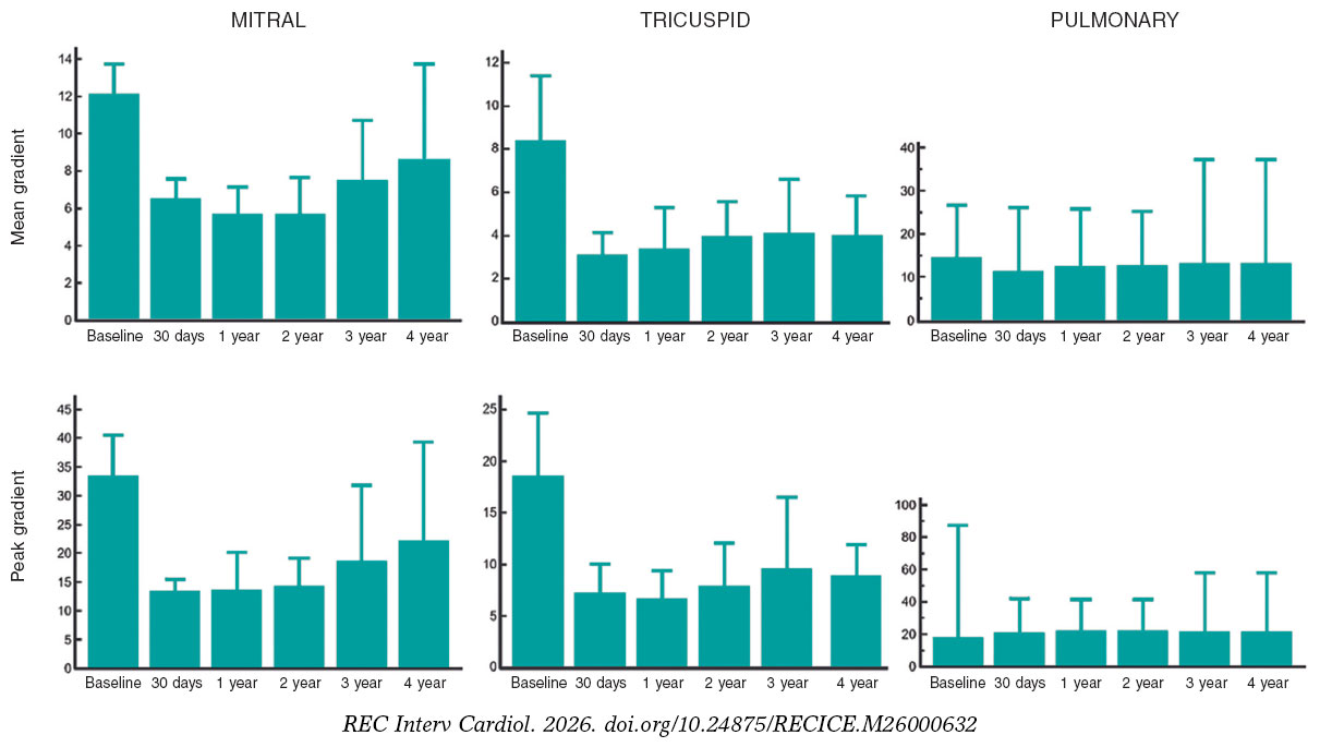
Figure 1. Changes in mean and peak transvalvular gradient over 4 years of follow-up after transcatheter mitral, tricuspid and pulmonary valve procedures.
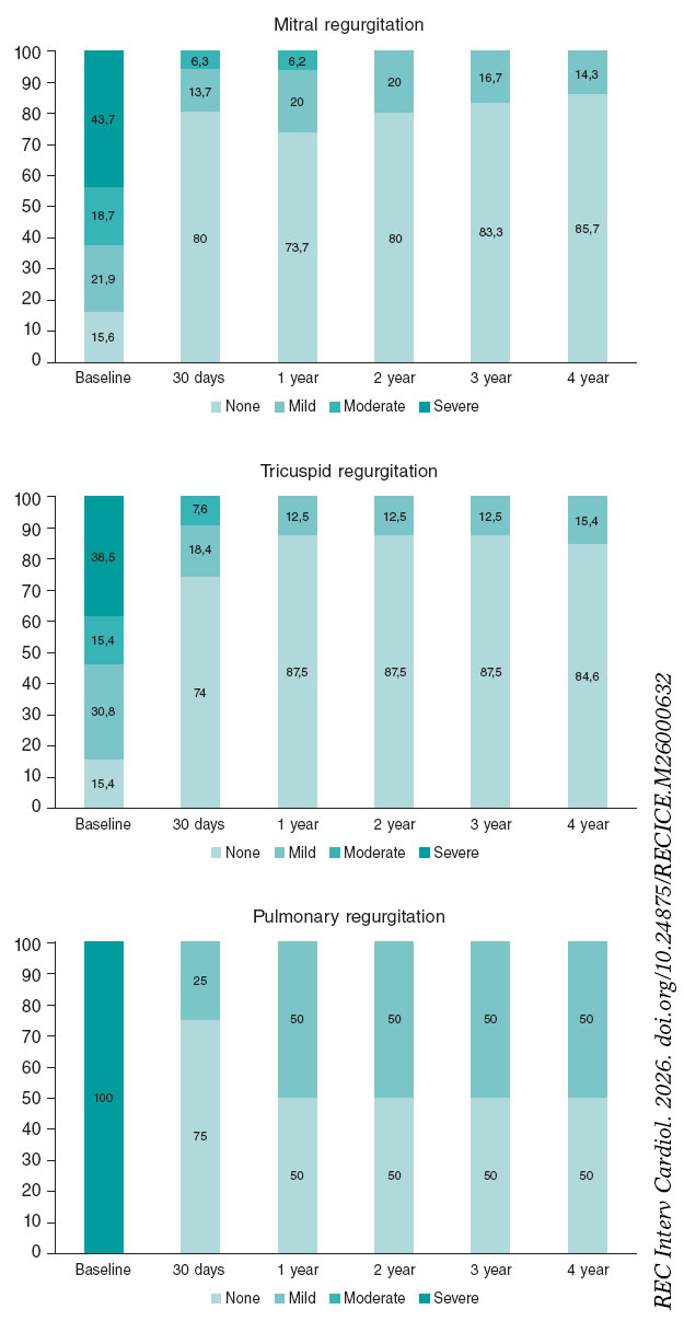
Figure 2. Changes in valvular regurgitation severity over 4 years of follow-up after transcatheter mitral, tricuspid and pulmonary valve procedures.
Survival and clinical outcomes
Clinical outcomes were assessed at the predefined 1- and 4-year time points. At 1 year, death had occurred in 2/33 (6.1%) mitral patients and in none of the tricuspid or pulmonary patients. No stroke was recorded in any group at 1 year. At 4 years, death had occurred in 4/33 (12.1%) mitral patients, 2/13 (15.3%) tricuspid patients, and 0/4 pulmonary patients. Stroke had occurred in 0/33 mitral, 1/13 (7.6%) tricuspid, and 0/4 pulmonary patients (results are presented including the confidence intervals for major estimates in table 3). No valve reintervention was recorded in any group at the 4-year follow-up.
Mitral procedural subgroups
Given the heterogeneity of the mitral cohort, outcomes were additionally explored according to procedural substrate (ViV, ViR, and ViMAC). Baseline, procedural, and follow-up characteristics of these subgroups are summarized in table S1. The operative risk was higher for patients undergoing ViMAC (11.5 ± 8.2) than for ViV (7.1 ± 5.4) or ViR (8.2 ± 6.8). Given the limited number of patients in each subgroup, no formal statistical comparisons were performed.
DISCUSSION
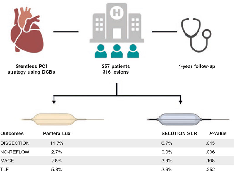
To our knowledge, this is the largest reported multicenter cohort with 4-year follow-up for Myval THV in non-aortic positions1 (figure 3). The present study provides descriptive long-term clinical and echocardiographic data on the use of this platform across 3 different non-aortic anatomical settings. Importantly, these findings should not be interpreted as a direct comparison with other balloon-expandable valves or as definitive evidence of valve durability. The main findings of this analysis are as follows: a) patients undergoing Myval implantation in non-aortic positions showed acceptable clinical outcomes at the 4-year follow-up, although estimates were imprecise given the limited sample size; b) among patients alive at 4 years, echocardiographic assessment showed preserved valve function, with no moderate or greater bioprosthetic regurgitation and no valve reinterventions; and c) stroke was a rare finding, with only 1 event recorded during follow-up.
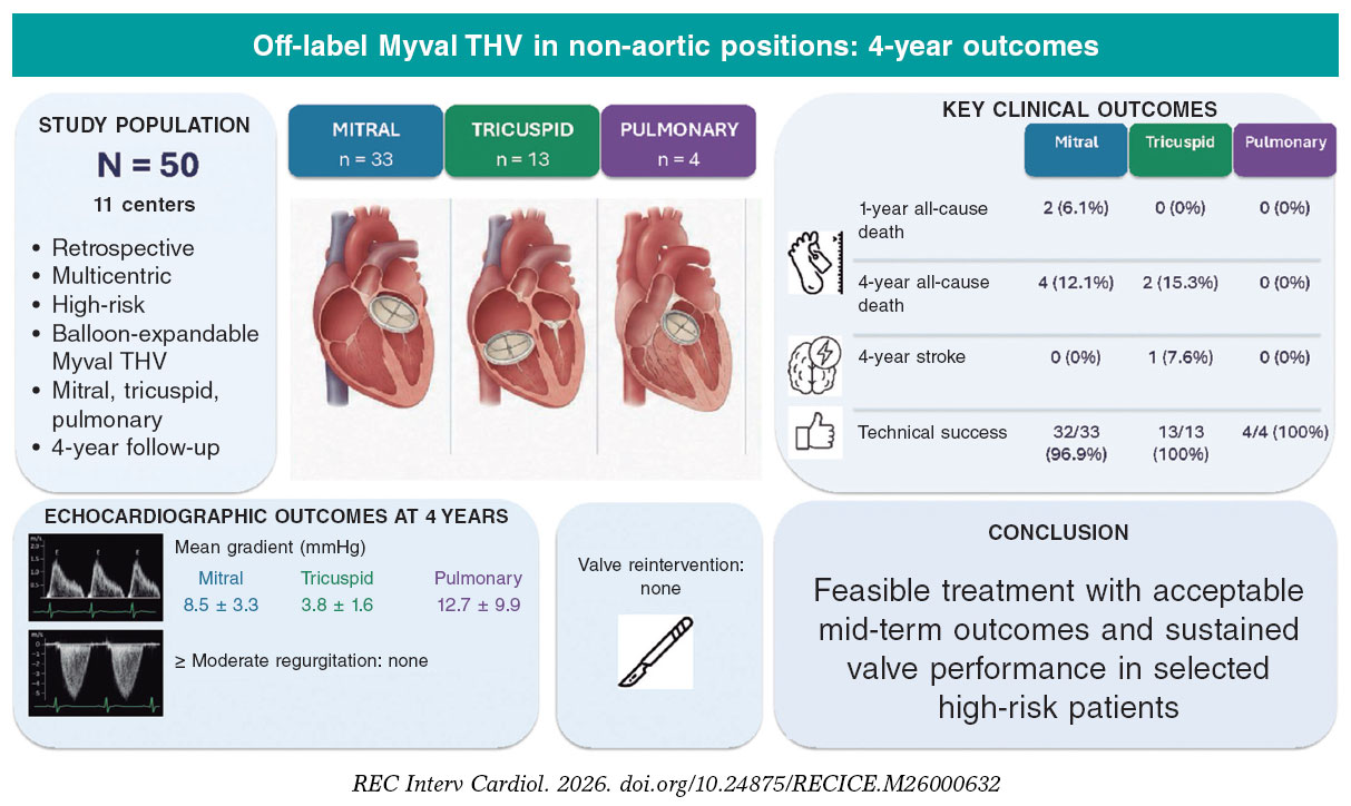
Figure 3. Central illustration. Clinical and echocardiographic outcomes after Myval implantation in the mitral, tricuspid, and pulmonary positions in a multicenter cohort of 50 high-risk patients. THV, transcatheter heart valve; mmHg, millimeters of mercury; N, total number of patients; n, number of patients in each subgroup.
Mitral procedures
Table 4 illustrates selected studies reporting mid- and long-term outcomes after mitral transcatheter procedures. An important aspect when interpreting these findings is differences in baseline patient characteristics and procedural substrate across studies. Our mitral cohort had a mean STS score of 7.9 ± 6.3%, and included ViV, ViR, and ViMAC procedures, which represent distinct anatomical and technical scenarios. For this reason, outcomes according to mitral substrate are additionally presented descriptively in table S1. A recent analysis of the intermediate-risk patients enrolled in the PARTNER 3 mitral valve-in-valve registry reported no deaths at the 1-year follow-up in 50 carefully selected patients, highlighting the favorable outcomes achievable with MViV when applied to patients without severe comorbidities.15 Another study showed higher 3-year survival after MViV in patients at low surgical risk as assessed by the STS score and in elective procedures whereas survival was significantly lower among patients with high STS scores and nonelective procedures.16 These studies provide useful clinical context but should not be directly compared with the present cohort because of differences in patient selection, procedural substrate, valve platform, and follow-up methodology. Our study showed a mean mitral gradient of 8.5 ± 3.3 mmHg at 4 years, compared with 5.53 ± 2.1 mmHg at 1 year. Although this numerical increase warrants attention, the limited sample size and descriptive design preclude determining whether it represents a meaningful longitudinal change. Importantly, no moderate or greater bioprosthetic regurgitation was identified at 4-year echocardiographic assessment, and no valve reintervention was recorded. Moreover, no prosthetic valve migration, endocarditis, or clinically diagnosed valve thrombosis was recorded during follow-up.
Table 4. Studies reporting mid- and long-term outcomes of transcatheter mitral ViV/ViR/ViMAC
| Procedure/device | Follow-up | Mean gradient (mmHg) | Survival (%) | Study |
|---|---|---|---|---|
| MViV – SAPIEN 3 | 1 year (up to 3 years registry) | 7 ± 2.9 | 83.3 (at 1 year) | Whisenant et al. (2020)20 |
| ViV/ViR/ViMAC – SAPIEN XT or SAPIEN 3 | 2 years | ViV 6.9 ± 2.4 |
ViV: 93.3 |
Eleid et al. (2022)21 |
| ViV/MViR/ViMAC – SAPIEN XT or SAPIEN 3 | 5 years | ViV: 6.6 ± 2.5 |
ViV: 78.6 |
Guerrero et al. (2023)22 |
| MViV- BEV | 5 years | 7.0 ± 2.4 (at 1 month) | 65 (at 5 years) | Schamroth et al. (2022)23 |
| MViV – SAPIEN 3 (intermediate-risk patients) | 1 year | 6.3 (at 1 year) | 100 | Malaisrie et al. (2024)15 |
|
Data are expressed as % or mean ± standard deviation. MViV, mitral valve-in-valve; ViMAC, valve-in-mitral annular calcification; ViR, valve-in-ring; ViV, valve-in-valve. |
||||
Tricuspid procedures
Data on transcatheter tricuspid procedures are limited. A midterm report from the VIVID registry of 306 patients undergoing tricuspid ViV or ViR reported cumulative 3-year incidences of death, reintervention, and valve-related adverse outcomes (endocarditis, thrombosis, or significant dysfunction) of 17%, 12%, and 8%, respectively.4 Although our study reports a mortality rate of 15.4% at 4 years which is consistent with these findings, the small sample size and wide confidence interval preclude meaningful comparison with the VIVID registry. Mussayev et al. (2023) reported 5 cases of Myval implantation in dysfunctional tricuspid prostheses or rings, with 80% survival rates and no cases of valve failure at the 1-year follow-up.17 Another study reported 4 patients who underwent transcatheter tricuspid ViV using balloon-expandable Edwards SAPIEN XT and SAPIEN 3 valves. At a median follow-up of 32 months (range, 9-47 months) all patients were alive and had NYHA functional class I-II. Echocardiographic assessment at the long-term follow-up showed low transvalvular gradients and no significant valvular regurgitation in all cases.18 Another study reported tricuspid ViR implantation in 20 patients using either a SAPIEN valve or Melody valve (Medtronic, United States). No procedural deaths occurred. During a median follow-up of 12 months, 1 patient died and 2 underwent repeat transcatheter tricuspid ViR implantation, 1 of whom subsequently required surgical valve replacement.19 Overall, published data on long-term outcomes of transcatheter valve implantation in the tricuspid position remains limited. In our cohort, the mean transvalvular gradient was 3.8 ± 1.6 mmHg at 4 years, with no moderate or greater bioprosthetic regurgitation or valve reinterventions. Although these findings suggest sustained echocardiographic valve performance among surviving patients, the small cohort precludes firm conclusions regarding long-term valve durability.4,17-19
Pulmonary procedures
Several studies have reported mid- and long-term outcomes of transcatheter valve implantation in the pulmonary position (table 5).5-10 Studies evaluating Myval by Al Nasef et al.9 and Sivaprakasam et al.10 reported favorable early outcomes with the Myval THV. Our study, which included only 4 patients undergoing pulmonary valve implantaion, should be considered exploratory. No deaths, cases of infective endocarditis, valve reinterventions, or moderate or greater bioprosthetic regurgitation were recorded through 4 years of follow-up. The mean transvalvular gradient at the 4-year assessment was 12.7 ± 9.9 mmHg. However, the very small sample size introduces substantial statistical imprecision and precludes definitive conclusions regarding clinical outcomes or valve durability. The valve’s radial strength and flexibility may be particularly advantageous in accommodating the often irregular anatomy of the right ventricular outflow tract.
Table 5. Studies reporting mid- and long-term outcomes of transcatheter pulmonary valve intervention
| Valve | Follow-up duration | RVOT gradient (mmHg) | PR | Survival | Study |
|---|---|---|---|---|---|
| Medtronic Melody | Up to 10 years (median, 8.4 years) | 72% none/trivial, 26% mild, 3% moderate at 10 years | 90% at 10 years | Jones et al. (2022)5 | |
| Edwards SAPIEN | Median, 3.3 years | Peak gradient, 20 [16-28] | Moderate-to-severe PR: 3% at 1 year | incidence rate of cardiac death (0.40 per 100 patient-years) | Belahnech et al. (2025)6 |
| Edwards SAPIEN XT | 3 years | Peak gradient, 17.8 ± 12.4 | Mild or less PR in 91.2% | 98.4% at 3 years | Kenny et al. (2018)7 |
| Mean gradient, 10.2 ± 7.8 | |||||
| Alterra Adaptive Prestent and SAPIEN 3 THV | 6 months | Mean gradient, 9.0 [6.8-14.3] | No PR (53.3%); mild PR (46.7%) no moderate or severe PR at 6 months | 100% at 6 months | Shahanavaz et al. (2020)8 |
| Meril Myval | 360 days [164-525] | Peak gradient 10 mmHg post-implant | Moderate PR (5.6%) | No death reported | Al Nasef et al. (2024)9 |
| Meril Myval | 16 months [10-22] | 12.5 ± 5.8 | No PR at 16 months | 100% at 16 months | Sivaprakasam et al. (2021)10 |
|
PR, pulmonary regurgitation; RVOT, right ventricular outflow tract; THV, transcatheter heart valve. |
|||||
Overall, these findings contribute to the limited body of evidence on the use of the Myval platform in non-aortic positions providing clinical and echocardiographic outcomes through 4 years of follow-up in the mitral, tricuspid, and pulmonary positions.20-23 Importantly, these 3 groups should not be considered a homogeneous cohort, given their markedly different anatomy, anchoring mechanisms, loading conditions, procedural indications, and anticipated mechanisms of valve failure. Accordingly, the present findings are primarily descriptive and hypothesis-generating. Although they may provide useful insights for operators considering this off-label strategy in selected patients, they do not establish comparative performance compared with other balloon-expandable valves or provide evidence of long-term durability.
Limitations
This study has several important limitations. Its retrospective observational design, absence of a control group, and lack of independent event adjudication or central echocardiographic core-laboratory assessment limit the strength of the conclusions. Although consecutive patients undergoing Myval implantation were included, patients who were evaluated but ultimately did not receive Myval were not systematically recorded. Consequently, the total number of patients screened and the reasons for exclusion could not be reconstructed, and selection bias cannot be excluded.
Follow-up imaging was performed according to local clinical practice rather than a prospectively defined protocol. Although echocardiographic follow-up was completed among patients surviving to 4 years, missing examinations at earlier time points may have introduced attrition bias. Furthermore, restricting late assessment to survivors may have led to an underestimation of valve dysfunction due to survivorship bias. Neither imputation nor sensitivity analysis were performed to address missing echocardiographic data. In addition, the absence of systematic CT surveillance precluded assessment of subclinical leaflet abnormalities or thrombosis.
Exact dates of death and stroke were unavailable for all events; Consequently, clinical outcomes were assessed at predefined 1- and 4-year time points and time-to-event analyses could not be performed.
Finally, the cohort was small and heterogeneous, encompassing different anatomical positions and procedural substrates. The mitral cohort included ViV, ViR, and ViMAC procedures, whereas the pulmonary subgroup comprised only 4 patients. Accordingly, subgroup estimates are imprecise and should be considered exploratory. Moreover, MVARC definitions have not been specifically validated for tricuspid or pulmonary interventions. Collectively, selection, missing imaging data, and survivorship bias may have resulted in overly favorable estimates of long-term valve performance, although the magnitude of these potential biases cannot be quantified.
CONCLUSIONS
In this retrospective multicenter series of selected patients undergoing off-label Myval THV implantation in non-aortic positions, clinical and echocardiographic follow-up through 4 years demonstrated sustained valve performance among surviving patients, with no moderate or greater bioprosthetic regurgitation or valve reinterventions at 4 years. Although these findings provide preliminary support for the feasibility of this approach, they do not establish definitive evidence of long-term valve durability or comparative performance. Larger prospective studies with systematic imaging follow-up, standardized position- specific endpoint definitions, and independent event adjudication are needed to validate these findings.
FUNDING
None declared.
ETHICAL CONSIDERATIONS
The study was conducted in full compliance with the Declaration of Helsinki and was approved by the local Clinical Research Ethics Committee (CEIC) of the leading institution. Due to the retrospective design of the study, the requirement for specific informed consent for study participation was waived by the Ethics Committee. Informed consent for the clinical procedure was obtained according to standard institutional practice. SAGER guidelines have been followed with respect to possible sex/gender bias.
STATEMENT ON THE USE OF ARTIFICIAL INTELLIGENCE
No artificial intelligence tools were used while preparing this manuscript.
AUTHORS’ CONTRIBUTIONS
I.J. Amat-Santos and A. Jain designed the study, collected the data, and wrote the final version of the manuscript. The remaining authors helped gather data, critically reviewing the manuscript and approving the final version.
CONFLICTS OF INTEREST
I.J. Amat-Santos serves as a proctor for Meril Life. The institution received a unrestricted grant for the LANDMARK trial. J. Jose is a proctor for Meril Life. M. Montenegro da Costa is a proctor for Meril Life. M. Montorfano has received consultant fees from Abbott, Boston Scientific, Kardia, and Medtronic. S. Gunasekaran is a proctor for Myval and ACURATE neo2 and has received honoraria from Meril Life and Boston Scientific. A. Seth is a proctor for Myval, ACURATE neo2 and Sentinel Devices, principal investigator of the Myval-1 study and has received speaker honoraria from Meril Life and Boston Scientific. A. Jain has received consultant fees from Meril Life.
WHAT IS KNOWN ABOUT THE TOPIC?
- Balloon-expandable transcatheter heart valves are increasingly used off-label in mitral, tricuspid, and pulmonary positions as alternatives to high-risk redo surgery.
- Although early outcomes with the Myval transcatheter heart valve are promising, evidence on its long-term performance in non-aortic positions remains limited.
WHAT DOES THIS STUDY ADD?
- This multicenter study evaluated 4-year clinical and echocardiographic outcomes following Myval implantation in 50 patients across the mitral, tricuspid, and pulmonary positions.
- High procedural success and sustained valve performance were observed, with no moderate or severe bioprosthetic regurgitation at 4 years and no valve reinterventions throughout follow-up.
- These findings support the feasibility of Myval implantation in selected patients requiring non-aortic valve procedures, although larger prospective studies are needed to confirm long-term valve durability.
SUPPLEMENTARY DATA

Vídeo 1. Jain A. DOI: 10.24875/RECICE.M26000632
Vídeo 2. Jain A. DOI: 10.24875/RECICE.M26000632
Vídeo 3. Jain A. DOI: 10.24875/RECICE.M26000632
REFERENCES
1. García-Gómez M, Fernández-Cordón C, González-Gutiérrez JC, et al. The novel balloon-expandable Myval transcatheter heart valve:systematic review of aortic, mitral, tricuspid and pulmonary indications. Rev Esp Cardiol. 2025;78:447-464.
2. Regueiro A, Granada JF, Dagenais F, Rodés-Cabau J. Transcatheter Mitral Valve Replacement:Insights From Early Clinical Experience and Future Challenges. J Am Coll Cardiol. 2017;69:2175-2192.
3. Little SH, Bapat V, Blanke P, Guerrero M, Rajagopal V, Siegel R. Imaging Guidance for Transcatheter Mitral Valve Intervention on Prosthetic Valves, Rings, and Annular Calcification. JACC Cardiovasc Imaging. 2021;14:22-40.
4. McElhinney DB, Aboulhosn JA, Dvir D, et al.;VIVID Registry. Mid-Term Valve-Related Outcomes After Transcatheter Tricuspid Valve-in-Valve or Valve-in-Ring Replacement. J Am Coll Cardiol. 2019;73:148-157.
5. Jones TK, McElhinney DB, Vincent JA, et al. LJ. Long-Term Outcomes After Melody Transcatheter Pulmonary Valve Replacement in the US Investigational Device Exemption Trial. Circ Cardiovasc Interv. 2022;15:e010852.
6. Belahnech Y, Martí-Aguasca G, Dos-SubiràL, et al. Mid-term outcomes of percutaneous pulmonary valve replacement with Edwards-Sapien bioprosthesis in native right ventricular outflow tract. Sci Rep. 2025;15:3977.
7. Kenny D, Rhodes JF, Fleming GA, et al. 3-Year Outcomes of the Edwards SAPIEN Transcatheter Heart Valve for Conduit Failure in the Pulmonary Position From the COMPASSION Multicenter Clinical Trial. JACC Cardiovasc Interv. 2018;11:1920-1929.
8. Shahanavaz S, Balzer D, Babaliaros V, et al. Alterra Adaptive Prestent and SAPIEN 3 THV for Congenital Pulmonic Valve Dysfunction:An Early Feasibility Study. JACC Cardiovasc Interv. 2020;13:2510-2524.
9. Al Nasef M, Erdem A, Aldudak B, et al. Multicenter Experience for Early and Mid-Term Outcome of MyVal Transcatheter Pulmonary Valve Implantation. Pediatr Cardiol. 2024;45:570-579.
10. Sivaprakasam MC, Reddy JRV, Gunasekaran S, et al. Early multicenter experience of a new balloon expandable MyVal transcatheter heart valve in dysfunctional stenosed right ventricular outflow tract conduits. Ann Pediatr Cardiol. 2021;14:293-301.
11. Vandenbroucke JP, von Elm E, Altman DG, et al.;STROBE Initiative. Strengthening the Reporting of Observational Studies in Epidemiology (STROBE):explanation and elaboration. Int J Surg. 2014;12:1500-1524.
12. Sharma SK, Rao RS, Chandra P, et al.;Collaborators. First-in-human evaluation of a novel balloon-expandable transcatheter heart valve in patients with severe symptomatic native aortic stenosis:the MyVal-1 study. EuroIntervention. 2020;16:421-429.
13. Baumbach A, van Royen N, Amat-Santos IJ, et al.;LANDMARK trial investigators. LANDMARK comparison of early outcomes of newer-generation Myval transcatheter heart valve series with contemporary valves (Sapien and Evolut) in real-world individuals with severe symptomatic native aortic stenosis:a randomised non-inferiority trial. Lancet. 2024;403:2695-2708.
14. Stone GW, Adams DH, Abraham WT, et al.;Mitral Valve Academic Research Consortium (MVARC). Clinical Trial Design Principles and Endpoint Definitions for Transcatheter Mitral Valve Repair and Replacement:Part 2:Endpoint Definitions:A Consensus Document From the Mitral Valve Academic Research Consortium. J Am Coll Cardiol. 2015;66:308-321.
15. Malaisrie SC, Guerrero M, Davidson C, et al.;PARTNER 3 Mitral Valve-in-Valve Study Investigators. One-Year Outcomes of Transseptal Mitral Valve-in-Valve in Intermediate Surgical Risk patients. Circ Cardiovasc Interv. 2024;17:e013782.
16. Eleid MF, Krishnaswamy A, Kapadia S, et al. 3-Year Outcomes of Mitral Valve-in-Valve Therapy Using Balloon-Expandable Transcatheter Valves in the United States. JACC Cardiovasc Interv. 2025;18:1454-1466.
17. Mussayev A, Alimbayev S, Tanaliev N, et al. Case report:Transcatheter tricuspid valve-in-valve implantation using novel balloon-expandable aortic valve with 1 year follow-up. Front Cardiovasc Med. 2023;10:1152280.
18. Scarsini R, Lunardi M, Pesarini G, et al. Long-term follow-up after trans-catheter tricuspid valve-in-valve replacement with balloon-expandable aortic valves. Int J Cardiol. 2017;235:141-146.
19. Aboulhosn J, Cabalka AK, Levi DS, et al. Transcatheter Valve-in-Ring Implantation for the Treatment of Residual or Recurrent Tricuspid Valve Dysfunction After Prior Surgical Repair. JACC Cardiovasc Interv. 2017;10:53-63.
20. Whisenant B, Kapadia SR, Eleid MF, et al. One-Year Outcomes of Mitral Valve-in-Valve Using the SAPIEN 3 Transcatheter Heart Valve. JAMA Cardiol. 2020;5:1245-1252.
21. Eleid MF, Wang DD, Pursnani A, et al. 2-Year Outcomes of Transcatheter Mitral Valve Replacement in patients With Annular Calcification, Rings, and Bioprostheses. J Am Coll Cardiol. 2022;80:2171-2183.
22. Guerrero ME, Eleid MF, Wang DD, et al. 5-Year Prospective Evaluation of Mitral Valve-in-Valve, Valve-in-Ring, and Valve-in-MAC Outcomes:MITRAL Trial Final Results. JACC Cardiovasc Interv. 2023;16:2211-2227.
23. Schamroth Pravda N, Mishaev R, Levi A, et al. Five-Year Outcomes of patients With Mitral Structural Valve Deterioration Treated With Trans catheter Valve in Valve Implantation - A Single Center Prospective Registry. Front Cardiovasc Med. 2022;9:883242.