
ABSTRACT
Mechanical complications of acute myocardial infarction—ventricular septal rupture, papillary muscle rupture, and free-wall rupture—are rare (< 0.3%) but lethal, with in-hospital mortality rates of 40% to 90%. These conditions may cause cardiogenic shock and require management at high-volume surgical centers by a multidisciplinary heart team. Mechanical circulatory support (MCS), including intra-aortic balloon pump (IABP), Impella, and venoarterial extracorporeal membrane oxygenation (VA-ECMO) may serve as a bridge to hemodynamic stabilization before definitive surgical or percutaneous repair. IABP reduces afterload and shunt fraction, Impella provides active left ventricular unloading, and VA-ECMO provides full cardiopulmonary support in refractory shock. The 2025 American College of Cardiology/American Heart Association guidelines recommend individualized use of short-term MCS (class IIa–IIb; level of evidence, B–C). Randomized trials of cardiogenic shock have excluded these patients; therefore, the available evidence is derived from registries and observational series subject to substantial selection bias. In postinfarction ventricular septal defect, IABP is generally considered first-line support, although VA-ECMO is often required. Delayed surgery (> 7 days) is associated with improved outcomes, although selection bias remains an important confounder. Papillary muscle rupture requires emergency surgery, with IABP used as a bridge; Impella should be used cautiously. Free-wall rupture also mandates emergency surgery; and VA-ECMO may serve as a bridge. MCS may provide time for stabilization and recovery, but no mortality benefit has been conclusively demonstrated. Optimal use requires expert multidisciplinary care.
Keywords: Cardiogenic shock. Mechanical complications. Mechanical circulatory support.
RESUMEN
Las complicaciones mecánicas del infarto agudo de miocardio, rotura del septo interventricular, rotura del músculo papilar y rotura de la pared libre, son raras (< 0,3%), pero letales, con una mortalidad hospitalaria del 40-90%. Estas afecciones cursan con shock cardiogénico y requieren tratamiento en centros con experiencia quirúrgica cardiaca y equipos multidisciplinarios. El soporte circulatorio mecánico (SCM)—balón de contrapulsación intraaórtico (BCIAo), Impella y oxigenador extracorpóreo de membrana venoarterial (ECMO-VA)—actúa como puente a la estabilización hemodinámica antes de la reparación definitiva. Las guías 2025 del American College of Cardiology y la American Heart Association recomiendan SCM individualizado (clase IIa-IIb, nivel de evidencia B-C). Los ensayos aleatorizados han excluido estas complicaciones; la evidencia procede tanto de registros como de series observacionales. En la rotura del septo interventricular, el BCIAo es de primera línea, aunque el ECMO-VA suele ser necesario; la cirugía diferida (> 7 días) mejora los resultados. La rotura del músculo papilar requiere cirugía urgente con BCIAo como puente. La rotura de la pared libre exige cirugía urgente; el ECMO-VA puede ser el tratamiento puente. El SCM gana tiempo pero no se ha demostrado su beneficio en cuanto a mortalidad.
Palabras clave: Shock cardiogénico. Complicaciones mecánicas. Soporte circulatorio mecánico.
Abbreviations
AMI: acute myocardial infarction. IABP: intra-aortic balloon pump. LV: left ventricular. MCS: mechanical circulatory support. PIVSD: postinfarction ventricular septal defect. PMR: papillary muscle rupture. VA-ECMO: venoarterial extracorporeal membrane oxygenation. VSR: ventricular septal rupture.
INTRODUCTION
Mechanical complications of acute myocardial infarction (AMI)—ventricular septal rupture (VSR), papillary muscle rupture (PMR) causing acute mitral regurgitation, and free wall rupture—are rare but catastrophic events that typically occurr 3 to 7 days after AMI. They present with acute heart failure, cardiogenic shock, a new murmur, or hemodynamic collapse, and are associated with mortality rates of 40% to 90%, depending on the lesion type, severity of shock, and diagnostic delay.1-3
Despite advances in reperfusion, their incidence has declined to < 0.3% of AMI cases; however, mortality remains disproportionately high (in-hospital mortality for VSR and PMR: 40%–80%; mortality for free-wall rupture without surgery, nearly 100%).1-3 Early recognition using echocardiography is critical. Both the European Society of Cardiology and the American College of Cardiology guidelines recommend management at centers with cardiac surgical expertise and heart teams.4,5 Surgical repair is the definitive treatment, but operative mortality remains high (30%–50%), particularly among patients with shock or multiorgan dysfunction. Delayed surgery (> 7 days) may improve outcomes by allowing maturation of the infarcted tissue, although the apparent survival benefit is confounded by immortal time bias and patient selection.
The central therapeutic challenge is to achieve sufficient hemodynamic stabilization to permit definitive intervention without incurring device-related complications. Mechanical circulatory support (MCS) devices—including intra-aortic balloon pump (IABP), Impella, and venoarterial extracorporeal membrane oxygenation (VA-ECMO)—are the main tools for this purpose (figure 1).1,3,6
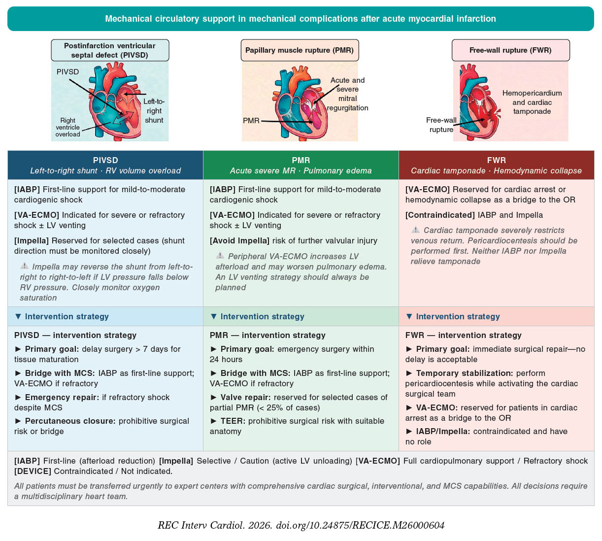
Figure 1. Central illustration. Decision algorithm for selecting MCS stratified by the type of mechanical complication and severity of cardiogenic shock. All patients should be transferred urgently to high-complexity centers with comprehensive cardiac surgical, interventional cardiology, and MCS capabilities. Evidence basis: 2025 American College of Cardiology/American Heart Association guidelines (Class IIa–IIb recommendations, Level of Evidence B–C); Ronco et al. 20217 (PIVSD systematic review, n = 2440); Kettner et al. 20138 (IABP in VSR/PMR); Matteucci et al. 20219 (FWR surgical outcomes). FWR, free-wall rupture; IABP, intra-aortic balloon pump; LV, left ventricular; MCS, mechanical circulatory support; MR, mitral regurgitation; OR, operating room; PIVSD, postinfarction ventricular septal defect; PMR, papillary muscle rupture; RV, right ventricular; TEER, transcatheter edge-to-edge repair; VA-ECMO, venoarterial extracorporeal membrane oxygenation.
In mild-to-moderate shock, IABP may provide initial stabilization; in severe or refractory shock, escalation to Impella or VA-ECMO may be required. The 3 complications differ substantially in their pathophysiology and hemodynamic effects, requiring individualized device selection5 (table 1).
Table 1. Summary of MCS device selection in mechanical complications of AMI
| Clinical scenario | PIVSD | PMR | FWR | ||
|---|---|---|---|---|---|
| Intervention | Surgical repair | Percutaneous closure | Surgery/MVR | TEER (selected) | Emergency surgery |
| Timing | Delayed (> 7 days) preferred; emergent if refractory shock | Bridge to surgery or alternative in patients at prohibitive surgical risk | Emergency (< 24 h) in most cases | Prohibitive surgical risk and suitable anatomy | Immediate surgery; pericardiocentesis only as temporizing measure |
| In-hospital mortality | 30%–50%12 | High; limited data13 | 18%–25%2 | Limited data17 | > 50% even with surgery20 |
| IABP role | [FIRST-LINE] Reduces SVR and left-to-right shunting (Qp:Qs reduced by 22%)7,14 | [ADJUNCT] Provides supportive hemodynamic assistance | [FIRST-LINE] Reduces the regurgitant fraction and increases CO by approximately 0.5 L/min8 | [ADJUNCT] Provides supportive hemodynamic assistance | [NOT INDICATED] Does not address cardiac tamponade21 |
| Impella role | [SELECTIVE] Active LV unloading; risk of right-to-left shunting if LV pressure falls below RV pressure7 | [SELECTIVE] Requires close monitoring of shunt direction | [CAUTION] The inflow cannula may worsen MR or cause chordal injury18 | [CONTRAINDICATED] | [CONTRAINDICATED] May increase bleeding through the rupture site21 |
| VA-ECMO role | [REFRACTORY SHOCK] Provides full cardiopulmonary support; LV venting recommended7 | [SALVAGE] May serve as a bridge to definitive repair | [SALVAGE] May serve as a bridge to surgery but is associated with a high complication rate and 39% mortality19 | [NOT STANDARD] | [BRIDGE TO OR] Effectiveness may be limited by impaired venous return due to tamponade; independent predictor of operative mortality9 |
| MCS complications | Hemolysis, limb ischemia (up to 12.5%), bleeding, AKI; risk increases with support duration11 | Bleeding, limb ischemia, renal failure; Impella-related mitral valve injury10,18 | Bleeding, inadequate VA-ECMO flow; high operative mortality with preoperative VA-ECMO9 | ||
| Key evidence | Ronco et al.7 2021 Kettner et al.8 2013 Ronco et al.15 2023 | Giblett et al.13 2020 | Massimi et al.16 2025 Kettner et al.8 2013 | Haberman et al.17 2025 | Matteucci et al.9 2021 |
|
AKI, acute kidney injury; AMI, acute myocardial infarction; CO, cardiac output; ECMO, extracorporeal membrane oxygenation; FWR, free-wall rupture; IABP, intra-aortic balloon pump; LV, left ventricular; MCS, mechanical circulatory support; MR, mitral regurgitation; MVR, mitral valve replacement; OR, operating room; PIVSD, postinfarction ventricular septal defect; PMR, papillary muscle rupture; Qp,Qs, pulmonary-to-systemic flow ratio; RV, right ventricular; SVR, systemic vascular resistance; TEER, transcatheter edge-to-edge repair; VA-ECMO, venoarterial extracorporeal membrane oxygenation. Badge colours: [FIRST-LINE] green = first-line; [SELECTIVE/CAUTION] orange = selective or caution; [SALVAGE] purple = refractory shock/salvage; [CONTRAINDICATED] red = contraindicated or not indicated. |
|||||
Prolonged MCS is associated with time-dependent increases in major complications including bleeding (30%–50%), limb ischemia, renal failure, stroke, and hemolysis. Complication rates increase with the duration of support and may influence the timing of definitive intervention.10,11
Postinfarction ventricular septal defect
Postinfarction ventricular septal defect (PIVSD) occurs in approximately 0.2% to 0.3% of AMI cases. Without repair, 30-day mortality approaches 80%; overall surgical mortality for all-comers remains 30%–50% and is among patients undergoing emergency repair in the setting of shock.22
The underlying pathophysiological mechanism is full-thickness septal necrosis with an abrupt left-to-right shunt, resulting in acute right ventricular volume overload, reduced systemic cardiac output, and pulmonary edema. Anterior or apical defects predominate, whereas inferior or posterior defects are more complex and are frequently accompanied by ischemic mitral regurgitation and right ventricular dysfunction.7,22
Patients typically present with a new, harsh systolic murmur and rapid hemodynamic deterioration. Diagnosis relies on emergency echocardiography to assess defect location and size, the pulmonary- to-systemic blood flow ratio (Qp:Qs), and biventricular function. Hemodynamic assessment demonstrates a step-up in oxygen saturation at the right ventricular level and elevated pulmonary capillary wedge pressure. Multimodality imaging, including transesophageal echocardiography, computed tomography, and cardiac magnetic resonance imaging, can further define the anatomy and inform the choice between surgical and transcatheter treatment. However, most patients are too unstable to undergo imaging beyond bedside echocardiography.23,24
Surgical repair remains the standard of care. Percutaneous closure is an alternative for patients at prohibitive surgical risk or may be used as a temporizing measure. Both approaches are associated with higher mortality when performed during the acute phase (< 7 days). Delayed surgery (> 7 days), which allows maturation of necrotic tissue, has been associated with better outcomes in observational studies. However, this association may reflect patient selection rather than a true treatment effect, because delayed surgery may be a marker of survival to intervention rather the benefit.12,13 Prolonged MCS as a bridge to definitive treatment carries substantial risks, including hemolysis requiring device removal, limb ischemia in up to 12.5% of patients undergoing femoral cannulation, and progressive organ failure. These complications frequently require earlier intervention than initially intended.11
Intra-aortic balloon pump
IABP support reduces systemic vascular resistance by approximately 15%, decreases the left-to-right shunt ratio (Qp:Qs) by 22% (eg, from 3.1 to 2.4), and increases systemic cardiac output by 12% (eg, from 2.1 L/min to 2.4 L/min) in PIVSD.14 Its mechanism—diastolic balloon inflation to augment coronary perfusion and presystolic deflation to reduce left ventricular (LV) afterload—directly addresses the hemodynamic consequences of VSR. IABP is the recommended first-line MCS device for hemodynamic stabilization in PIVSD.7,25
A single-center cohort found that IABP use in patients with shock was associated with lower 30-day mortality than no IABP (61% vs 100%), and all patients who did not undergo repair died within 3 days.8 However, the 100% mortality observed without IABP in small cohorts likely reflects selection bias and confounding by indication rather than proven IABP efficacy. A systematic review of 2440 patients with PIVSD found that 93% received IABP support and that in-hospital mortality was 52%.7 No randomized data are available specifically for PIVSD because the IABP-SHOCK II trial excluded patients with mechanical complications.26
IABP is often insufficient in patients with severe cardiogenic shock with multiorgan dysfunction. Escalation to higher-level MCS is required when instability persists because of the device’s limited effect on cardiac output.
Impella
Impella-mediated LV unloading reduces LV end-diastolic pressure and afterload, thereby lowering pulmonary capillary wedge pressure and the left-to-right shunt fraction.25 In the systematic review by Ronco et al., only 17 of 2240 patients with PIVSD received Impella support; the in-hospital mortality rate was 35%, and complications included hemolysis, stroke, and LV thrombus.7
An important caveat is that Impella may reduce LV pressure below right ventricular pressure, particularly at excessive pump flow or in the presence of large defects, thereby promoting right-to-left shunting, systemic desaturation, and cerebral or coronary hypoxia.7 Expert consensus therefore considers PIVSD a relative contraindication to Impella support. Impella may nevertheless be used selectively for temporary LV unloading as a bridge to definitive closure, with careful monitoring of shunt direction and hemodynamic response.
A retrospective case series of 4 patients showed that preoperative Impella 5.5 implantation performed under local anesthesia in 3 patients, permitted a mean time to surgery of 9.8 ± 3.1 days, and 75% of patinets were successfully weaned from support postoperatively.27
Venoarterial extracorporeal membrane oxygenation
VA-ECMO provides full cardiopulmonary support and is the only device capable of fully stabilizing patients with refractory cardiogenic shock or multiorgan failure complicating PIVSD.25 Peripheral VA-ECMO increases mean arterial pressure through retrograde aortic flow but also increases LV afterload, which may exacerbate pulmonary edema and potentially increase the left-to-right shunting. LV venting with an IABP or Impella is recommended in the presence of refractory pulmonary edema, increasing pulmonary capillary wedge pressure, absent aortic valve opening, or a spherically dilated LV on echocardiography.5,6,10
In the systematic review, 144 of 2440 patients received VA-ECMO as a bridge therapy to definitive treatment; the in-hospital mortality rate was 29%, and major complications including bleeding, limb ischemia, and coagulopathy.7 Small case series have demonstrated the feasibility of VA-ECMO as a bridge to delayed repair, typically performed after 5 to 14 days, with survival to hospital discharge in approximately one-half to two-thirds of patients who ultimately underwent repair.28-30
ECpella, consisting of combined VA-ECMO and Impella support, can unload the LV while providing full systemic circulatory support. Registry data indicate that ECpella is feasible but associated is with high complication rates and has not demonstrated lower in-hospital mortality compared with VA-ECMO plus IABP or VA-ECMO alone.3 The European survey by Ronco et al. documented substantial heterogeneity in current management strategies.15
PAPILLARY MUSCLE RUPTURE
PMR complicates < 0.1% of AMI cases but is associated with an in-hospital mortality rate of 20% to 40% even with surgical intervention, and 80% without surgery.2,3,6,31 The posteromedial papillary muscle is more frequently affected because it typically receives its blood supply solely from the posterior descending artery. Complete rupture causes immediate hemodynamic collapse, whereas partial rupture may permit transient stabilization.
The pathophysiological mechanism is acute, severe mitral regurgitation causing sudden volume overload of a normal-sized LV and left atrium, leading to rapid-onset pulmonary edema and reduced cardiac output. Unlike in chronic mitral regurgitation, these chambers cannot accommodate the abrupt increase in volume, resulting in a fulminant clinical presentation.
Diagnosis relies on echocardiography, which may demonstrate a flail mitral leaflet, severe mitral regurgitation, and direct visualization of the ruptured papillary muscle. Mitral valve replacement is the standard treatment. Mitral valve repair may be feasible in selected patients with partial PMR cases but is associated with higher recurrence rates.16 Emergent surgery within 24 hours is recommended. In patients at prohibitive surgical risk, transcatheter edge-to-edge repair may be considered when the anatomy is suitable.17
Intra-aortic balloon pump
IABP is the recommended first-line MCS for PMR. Afterload reduction decreases the regurgitant fraction across the mitral valve and improves forward cardiac output. A cohort study found IABP use was associated with lower 30-day mortality (61% vs 100%; P = .04) and lower preoperative mortality (11% vs 88%; P < .001) compared with no IABP use among patients with acute mitral regurgitation and cardiogenic shock.8 These comparisons were observational and subject to selection bias; nevertheless, IABP remains recommended because its mechanism directly reducies regurgitant volume.
Impella
Impella should be used cautiously in patients with PMR. The inflow cannula may directly traumatize the mitral valve apparatus, including the chordae thendineae or papillary muscle structures, potentially worsening regurgitation.18 In the setting of PMR with mobile or detached fragments, the risk of further structural damage, device malfunction, or embolization is heightened. Evidence is limited to case series and expert opinion.1,2
Venoarterial extracorporeal membrane oxygenation
VA-ECMO can stabilize end-organ perfusion in refractory shock as a bridge to surgery. Observational multicenter data indicate VA-ECMO is used preoperatively in approximately 10% of surgically treated patients with PMR; however, in-hospital mortality remains high (39% vs 22% among surgical patients not receiving VA-ECMO).19,32 The poorer outcomes observed in the VA-ECMO group likely reflect greater baseline severity and higher complication rates, including bleeding, limb ischemia, renal failure. Peripheral cannulation increases LV afterload and may worsen pulmonary edema; therefore, LV venting strategies should be planned when VA-ECMO is initiated.
FREE-WALL RUPTURE
Free-wall rupture is a distinct clinical emergency requiring immediate surgical intervention. It differs fundamentally from PIVSD and PMR because no MCS device can substitute for surgical repair. It typically results from transmural infarction, causing hemopericardium and cardiac tamponade with rapid hemodynamic collapse. In-hospital mortality exceeds 50% even with surgery and approaches 100% without surgical treatment.20
Emergency surgery is the only potentially curative intervention. Pericardiocentesis may provide temporary relief of tamponade but must be followed by definitive surgical repair.3
Intra-aortic balloon pump and Impella
Neither IABP nor Impella has an established role in free-wall rupture. These 2 devices are designed to treat pump failure rather than structural disruption or cardiac tamponade. Impella is specifically contraindicated in LV rupture because it cannot provide effective support when ventricular chamber integrity is lost and may increase bleeding through the rupture site.21 IABP may have a very limited temporizing role in subacute or oozing ruptures but is ineffective in classic free-wall rupture with rapid hemodynamic collapse.
Venoarterial extracorporeal membrane oxygenation
VA-ECMO may be used as salvage therapy to maintain end-organ perfusion while emergency surgery is arranged, particularly in patients with cardiac arrest.33,34 A critical limitation in free-wall rupture is that cardiac tamponade severely restricts venous return, thereby limiting VA-ECMO flow and effectiveness. Inadequate venous drainage through the inferior vena cava cannula may render support partially or completely ineffective. Furthermore, a multicenter study identified preoperative VA-ECMO use in patients with free-wall rupture as an independent predictor of increased operative mortality.9 In patients presenting with cardiac arrest, VA-ECMO bedside cannulation may nevertheless represent the only means of temporary stabilization before emergency surgical repair.
DISCUSSION
Mechanical complications of AMI remain clinically relevant despite their declining incidence because the associated morbidity and mortality remain extremely high. Several key messages emerge from this review of the available evidence.
First, all patients with suspected mechanical complications should be transferred urgently to high-complexity centers with comprehensive cardiac surgery, interventional cardiology, advanced MCS, cardiac imaging, and intensive care capabilities. Multidisciplinary heart team decision-making is essential and has been associated with improved adherence to clinical guidelines and better outcomes in patiens with cardiogenic shock.1,5
Second, in severe refractory cardiogenic shock, VA-ECMO is the only device capable of providing full cardiopulmonary support irrespective of native cardiac function. IABP and Impella provide only partial support and may be insufficient in patients with multiorgan failure. The objective of MCS is to maintain end-organ perfusion as a bridge to definitive repair, not to replace it.
Third, in patients with PIVSD or PMR, appropriate MCS may permit delayed surgical intervention, which has been associated with improved outcomes in observational data. Although the apparent benefit of delayed surgery may partly reflect survivorship bias, there is a plausible biological rationale: maturation of necrotic tissue reduces intraoperative friability and may improve the durability of repair. Future trials should determine whether protocolized MCS-based bridging strategy improve outcomes beyond the effects of patient selection.12,13
Fourth, free-wall rupture is a fundamentally different emergency in which surgery is the only potentially lifesaving intervention. VA-ECMO may serve as a bridge in patients with cardiac arrest, although its effectiveness may be limited by impaired venous return due to cardiac tamponade. IABP and Impella have no established role.
The absence of randomized trial evidence in this population is a major limitation. Trials such as IABP-SHOCK II, DANGER-SHOCK, ECLS-SHOCK, and ECMO-CS excluded patients with mechanical complications.26,35-37 The European survey by Ronco et al.15 documented substantial heterogeneity in current management, underscoring the need for standardized treatment protocols.
Medical therapy alone is associated with near-universal early mortality. Vasoactive drugs should be used cautiously: dobutamine may worsen VSR progression, whereas noradrenaline may increase the severity of mitral regurgitation. Echocardiography, most often performed at the bedside because of patient instability, remains the principal diagnostic modality.
Future research priorities include: a) prospective registries incorporating causal inference methods; b) pragmatic registry-based trials and adaptive platform designs; c) definition of optimal patient selection and timing for surgical vs percutaneous repair; d) evaluation of emerging percutaneous treatments, including transcatheter edge-to-edge repair for PMR and transcatheter closure of VSR; e) development and validation of risk stratification tools; and f) investigation of the indications for cardiac transplantation and durable left ventricular assist devices.
CONCLUSIONS
Mechanical complications of AMI are life-threatening emergencies in which MCS may serve as a bridge to hemodynamic stabilization before definitive surgical or percutaneous repair. Device selection must be individualized according to the type of mechanical complication, severity of shock, and degree of organ dysfunction. IABP is the preferred first-line device for PIVSD and PMR; VA-ECMO provides the only full cardiopulmonary support in refractory shock; Impella should be used cautiously in both VSR because of the risk of right-to-left shunting and PMR because of the risk of mitral valve injury; and free-wall rupture is a surgical emergency in which IABP and Impella have no established role. Until high-quality evidence becomes available, the use of MCS should be guided by pathophysiological principles and multidisciplinary expertise rather than device-driven enthusiasm. All patients with these complications should be managed at expert centers with comprehensive cardiac surgical, interventional, and MCS capabilities.
FUNDING
None declared.
STATEMENT ON THE USE OF ARTIFICIAL INTELLIGENCE
During the preparation of this work, the authors used a generative artificial intelligence language model (ChatGPT, OpenAI) to assist solely with minor language editing and stylistic refinement of selected passages. All content was subsequently reviewed and edited by the authors, who take full responsibility for the final manuscript. The tool was not used for study design, data analysis, interpretation of results, or the generation of scientific content; these tasks were performed entirely by the authors.
AUTHORS’ CONTRIBUTIONS
R. Lorca and P. Avanzas shared senior authorship. All authors made substantial contributions to this work; participated in drafting the manuscript and critically revised it for important intellectual content; reviewed and approved the final version; and agreed to be accountable for all aspects of the work by ensuring that questions related to the accuracy or integrity of any part of the work are properly investigated and resolved.
CONFLICTS OF INTEREST
None declared.
REFERENCES
1. Murphy A, Goldberg S. Mechanical Complications of Myocardial Infarction. Am J Med. 2022;135:1401–1409.
2. Gong FF, Vaitenas I, Malaisrie SC, Maganti K. Mechanical Complications of Acute Myocardial Infarction. JAMA Cardiol. 2021;6:341.
3. Damluji AA, van Diepen S, Katz JN, et al. Mechanical Complications of Acute Myocardial Infarction: A Scientific Statement From the American Heart Association. Circulation. 2021;144:e16-e35.
4. Byrne RA, Rossello X, Coughlan JJ, et al. 2023 ESC Guidelines for the management of acute coronary syndromes. Eur Heart J. 2023;44: 3720–3826.
5. Rao SV, O'Donoghue ML, Ruel M, et al. 2025 ACC/AHA/ACEP/NAEMSP/SCAI Guideline for the Management of Patients With Acute Coronary Syndromes. J Am Coll Cardiol. 2025;85:2135–2237.
6. Saggu JS, Seelhammer TG, Esmaeilzadeh S, et al. Mechanical Circulatory Support for Acute Myocardial Infarction Cardiogenic Shock. J Cardiothorac Vasc Anesth. 2025;39:1049–1066.
7. Ronco D, Matteucci M, Ravaux JM, et al. MCS as Bridge in Post-Infarction VSR. JACC Cardiovasc Interv. 2021;14:1053–1066.
8. Kettner J, Sramko M, Holek M, Pirk J, Kautzner J. Utility of IABP for VSR and Acute MR. Am J Cardiol. 2013;112:1709–1713.
9. Matteucci M, Kowalewski M, De Bonis M, et al. Surgical Treatment of Post-Infarction LV Free-Wall Rupture. Ann Thorac Surg. 2021;112: 1186–1192.
10. Geller BJ, Sinha SS, Kapur NK, et al. Escalating and De-escalating Temporary Mechanical Circulatory Support in Cardiogenic Shock. Circulation. 2022;146:e50-e68.
11. Ott S, Germinario L, Müller-Wirtz LM, et al. Impact of complications on survival in temporary mechanical circulatory support. J Heart Lung Transplant. 2025;44:880–891.
12. Ronco D, Matteucci M, Kowalewski M, et al. Surgical Treatment of Postinfarction VSR. JAMA Netw Open. 2021;4:e2128309.
13. Giblett JP, Jenkins DP, Calvert PA. Transcatheter treatment of postinfarct VSD. Heart. 2020;106:878–884.
14. Testuz A, Roffi M, Bonvini RF. Left-to-right shunt reduction with IABP in post-MI VSD. Catheter Cardiovasc Interv. 2013;81:727–731.
15. Ronco D, Ariza-Solé A, Kowalewski M, et al. The current clinical practice for management of post-infarction ventricular septal rupture: a European survey. Eur Heart J Open. 2023;3:oead091.
16. Massimi G, Matteucci M, De Bonis M, et al. Repair Versus Replacement in PMR: A Multicenter Study. Eur J Cardiothorac Surg. 2025;67:ezaf284.
17. Haberman D, Estévez-Loureiro R, Czarnecki A, et al. TEER in severe MR following AMI. Eur J Heart Fail. 2025;27:912–921.
18. Sef D, Kabir T, Lees NJ, Stock U. Valvular complications following Impella implantation. J Card Surg. 2021;36:1062–1066.
19. Kowalewski M, Zielin´ski K, Brodie D, et al. VA-ECMO for Postcardiotomy Shock—ELSO Registry. Crit Care Med. 2021;49:1107–1117.
20. Antunes MJ. Left ventricular free wall rupture. J Card Surg. 2021;36: 3334–3336.
21. Bernhardt AM, Copeland H, Deswal A, et al. ISHLT/HFSA Guideline on Acute Mechanical Circulatory Support. J Card Fail. 2023;29:304–374.
22. Cubeddu RJ, Lorusso R, Ronco D, Matteucci M, Axline MS, Moreno PR. Ventricular Septal Rupture After Myocardial Infarction. J Am Coll Cardiol. 2024;83:1886–1901.
23. Baltodano-Arellano R, Cupe-Chacalcaje K, Cachicatari-Beltran A, et al. Echocardiography in VSR after AMI. Echocardiography. 2023;40: 1310–1324.
24. Dhaliwal S, Ducas R, Shuangbo L, et al. Multimodality imaging of VSR post MI. BMC Res Notes. 2012;5:583.
25. Shibasaki I, Otani N, Saito S, et al. Overview of MCS for post-MI VSR. J Cardiol. 2023;81:491–497.
26. Thiele H, Zeymer U, Neumann FJ, et al. Intraaortic Balloon Support for Myocardial Infarction with Cardiogenic Shock. N Engl J Med. 2012;367: 1287–1296.
27. Eghbalzadeh K, Wahlers TCW, Deppe AC. Implanting Impella 5.5 under Local Anesthesia. Thorac Cardiovasc Surg. 2024;72:296–299.
28. Rob D, Špunda R, Lindner J, et al. Rationale for early ECMO in postinfarction VSR with cardiogenic shock. Eur J Heart Fail. 2017;19(S2): 97–103.
29. Doi A, Negri JC, Marasco SF, et al. ECMO as Bridge to Repair of Postinfarct VSD. ASAIO Journal. 2023;69:101–106.
30. Watkins AC, Maassel NL, Ghoreishi M, et al. Preoperative VA-ECMO in Advanced Structural Heart Disease. Ann Thorac Surg. 2018;106: 1709–1715.
31. Samsky MD, Morrow DA, Proudfoot AG, et al. Cardiogenic Shock After Acute Myocardial Infarction. JAMA. 2021;326:1840.
32. Matteucci M, Fina D, Jiritano F, et al. ECMO in postinfarction mechanical complications: ELSO Registry. Interact Cardiovasc Thorac Surg. 2020;31:369–374.
33. Pineton de Chambrun M, Bréchot N, Combes A. VA-ECMO in cardiogenic shock. Curr Opin Crit Care. 2019;25:397–402.
34. Rao P, Khalpey Z, Smith R, Burkhoff D, Kociol RD. VA-ECMO for Cardiogenic Shock and Cardiac Arrest. Circ Heart Fail. 2018;11:e004905.
35. Ostadal P, Rokyta R, Karasek J, et al. ECMO-CS Randomized Clinical Trial. Circulation. 2023;147:454–464.
36. Thiele H, Zeymer U, Akin I, et al. Extracorporeal Life Support in Infarct- Related Cardiogenic Shock. N Engl J Med. 2023;389:1286–1297.
37. Møller JE, Engstrøm T, Jensen LO, et al. Microaxial Flow Pump or Standard Care in Infarct-Related Cardiogenic Shock. N Engl J Med. 2024;390: 1382–1393.