
This is the case of a 7-year-old girl weighing 19.3 kg with a diagnosis of Shone syndrome. She underwent neonatal surgery to close the ventricular septal defect and enlarge the aortic arch. At 20 months of age, she underwent subaortic membrane resection and mitral valve repair; and at 2 years of age, a Konno–Rastan procedure (anterior aortoventriculoplasty with implantation of a 16-mm ATS Open Pivot aortic heart valve [ATS Medical, Inc., United States]).
At the follow-up, we observed a paravalvular leak that progressed to hemodynamic significance, with a 4-mm vena contracta, left ventricular dilatation, and pandiastolic runoff in the thoracic aorta. Consequently, the transcatheter closure of the leak was planned.
Initial aortography did not demonstrate a paravalvular leak but revealed dilation of the left main coronary artery (6.5 mm) and a coronary–left ventricular fistula measuring 2.4 mm in diameter and 4.5 mm in length (figure 1 and video S1).
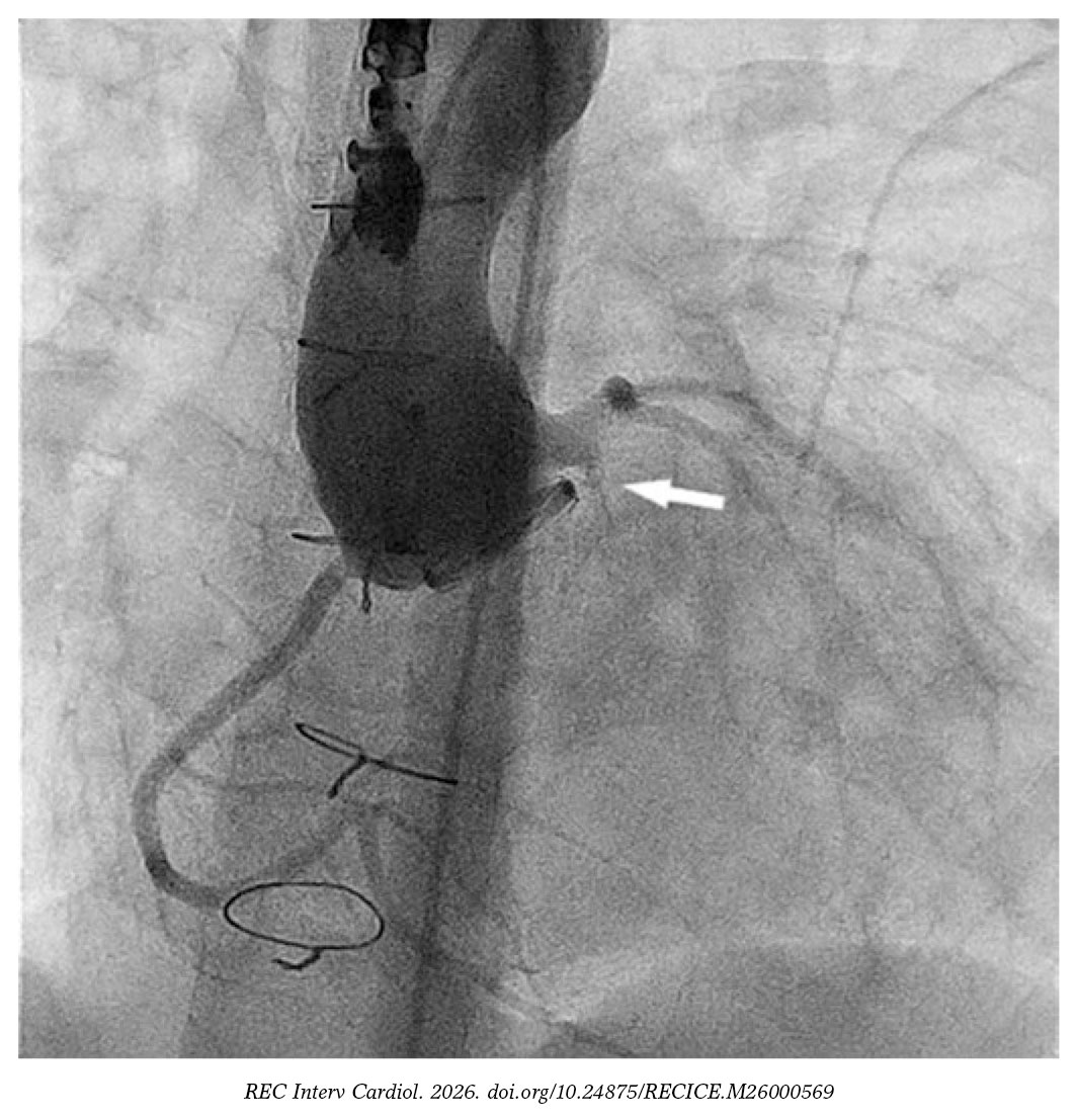
Figure 1.
We catheterized the fistulous tract using a modified 4-Fr NIH catheter (Cordis, United States) and a 0.035 in hydrophilic guidewire. Afterwards, we mounted a 4-Fr delivery catheter over the guidewire, and deployed a 4 mm × 4-mm Nit-Occlud PDA device (PFM Medical, Germany) (figure 2 and video S2). We chose this device over nitinol mesh occluders because their greater length and retention discs were considered at risk of interfering with the coronary artery.
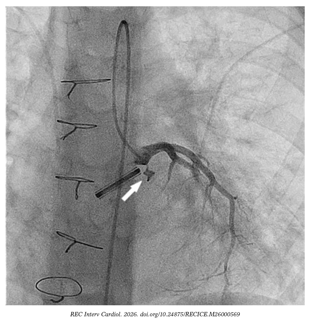
Figure 2.
Postoperative coronary fistulas may mimic paravalvular leaks. Their occlusion is recommended to prevent volume overload, progressive coronary artery dilatation, and the associated risk of myocardial ischemia.
FUNDING
None declared.
ETHICAL CONSIDERATIONS
As this is a case report involving a single patient, approval by an ethics committee was deemed unnecessary. Informed consent was obtained from the patient’s parents. The SAGER guidelines were not considered in the preparation of this case.
STATEMENT ON THE USE OF ARTIFICIAL INTELLIGENCE
No artificial intelligence was used in the preparation of this work.
AUTHORS’ CONTRIBUTIONS
All listed authors were directly involved in the drafting of this article, read and approved its final version.
CONFLICTS OF INTEREST
None declared.
SUPPLEMENTARY DATA
Vídeo 1. Santos Lorente C. DOI: 10.24875/RECICE.M26000569
Vídeo 2. Santos Lorente C. DOI: 10.24875/RECICE.M26000569


