
This is the case of an 86-year-old woman with symptomatic severe aortic stenosis. Prior to transcatheter aortic valve implantation (TAVI), coronary computed tomography angiography revealed the presence of an aortic annulus perimeter of 68 mm (figure 1A), severe valvular calcification of 2584 Agatston units (figure 1B), a 22 mm × 12 mm (figure 1D) soft plaque with an irregular surface in the descending thoracic aorta (figures 1C, E, arrows; figure 1F, ellipse), and adequate femoral accesses.
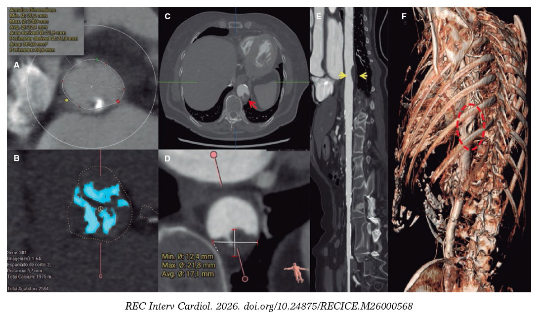
Figure 1.
To reduce the risk of aortic plaque-related complications during TAVI—particularly the risk of embolization (figure 2A, arrows)—we planned a staged strategy consisting of aortic stent-graft implantation followed by TAVI. Via a single 18-Fr transfemoral access, we implanted an 85 mm × 32 mm Endurant II covered stent-graft system (Medtronic, United States) (figure 2B-C and figure 2F, white asterisk). Afterwards, we advanced the TAVI delivery system through the stent-graft uneventfully (figure 2D, arrow), and implanted a 26-mm self-expanding Evolut FX+ valve (Medtronic, United States) (figure 2E-F, yellow asterisk), with no significant residual gradient and only mild paravalvular regurgitation on angiography. The patient was discharged without complications. Functional class improved, and transthoracic echocardiography confirmed the normal functioning of the TAVI at the 1-month follow-up.
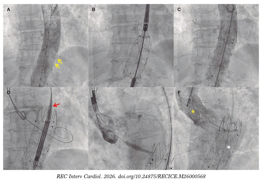
Figure 2.
This case illustrates the importance of multidisciplinary collaboration in the minimally invasive management of aortic disease to reduce complications and improve outcomes of transcatheter procedures.
Videos S1-S5 show the procedure and follow-up echocardiographic findings.
FUNDING
None declared.
ETHICAL CONSIDERATIONS
Written informed consent was obtained from the patient, and the study was conducted in full compliance with applicable ethical principles. SAGER guidelines were considered to minimize potential sex- and gender-related bias.
DECLARATION ON THE USE OF ARTIFICIAL INTELLIGENCE
No artificial intelligence or automated content generation tools were used in the preparation of this article.
AUTHORS’ CONTRIBUTIONS
M. García-Guimarães: conceptualization, methodology, and drafting of the original manuscript. J. M. Vegas-Valle, P. Miranda, and T. Guiberteau-Díaz: manuscript review and editing. M. J. Vallina-Victorero Vázquez and I. Lozano Martínez-Luengas: conceptualization, manuscript review, and editing.
CONFLICTS OF INTEREST
None declared.
SUPPLEMENTARY DATA
Vídeo 1. García-Guimarães M. DOI: 10.24875/RECICE.M26000568
Vídeo 2. García-Guimarães M. DOI: 10.24875/RECICE.M26000568
Vídeo 3. García-Guimarães M. DOI: 10.24875/RECICE.M26000568
Vídeo 4. García-Guimarães M. DOI: 10.24875/RECICE.M26000568
Vídeo 5. García-Guimarães M. DOI: 10.24875/RECICE.M26000568


