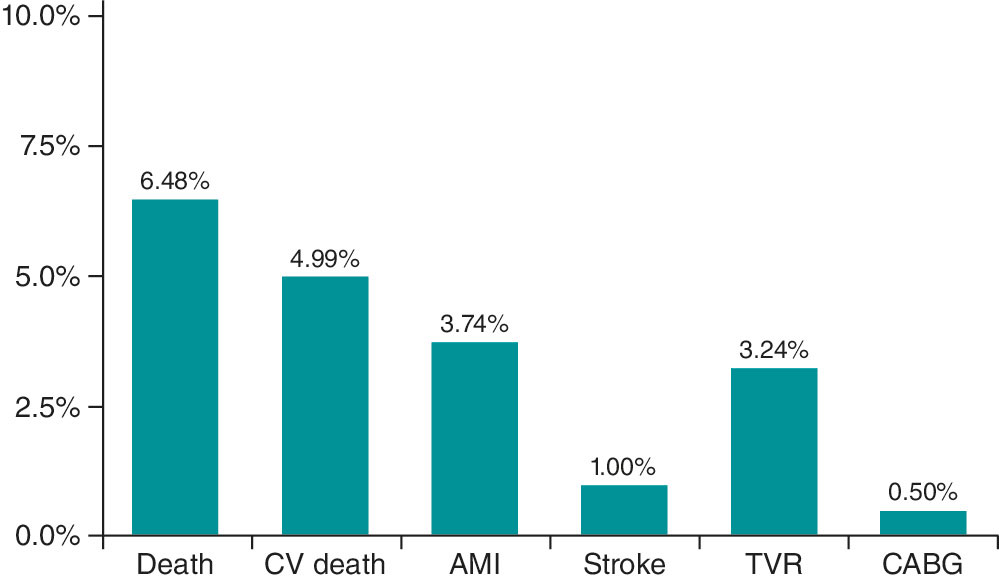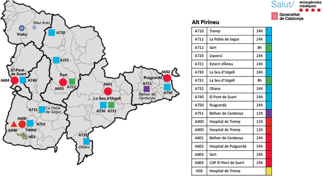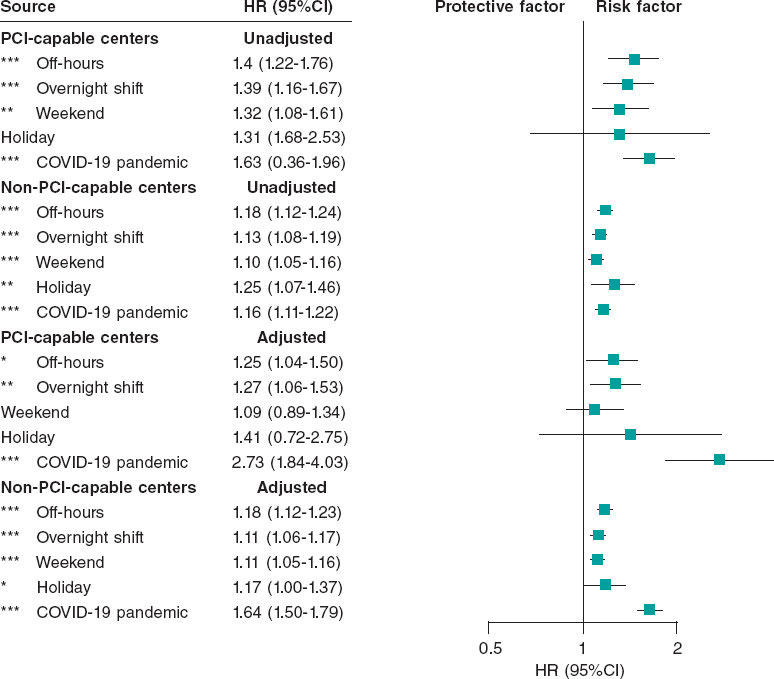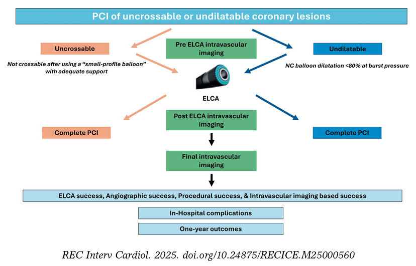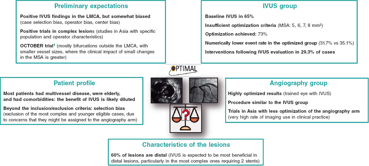


QUESTION: Data from two recent trials, OPTIMAL1 and IVUS-CHIP2, have yielded unexpected results. How do you interpret these studies?
ANSWER: The first conclusion one might draw from these two studies—to which ILUMIEN IV3 could be added, as it failed to show a clear overall clinical benefit too—is that the use of intravascular ultrasound in the percutaneous treatment of complex lesions or left main coronary artery disease does not improve patient outcomes compared with angiography-guided treatment alone. However, to be fair, this statement requires some qualification. I would add the terms “systematic use” and “in the hands of operators experienced in intracoronary imaging.” Both considerations are relevant because they underscore the importance of determining when intracoronary imaging is truly needed and ensuring that complex procedures are performed by appropriately experienced operators.
Q: Observational registries in left main coronary artery disease and randomized Asian trials in other complex lesions showed favorable outcomes with imaging. How can the discrepancy between those studies and the more recent trials be explained?
A: Intracoronary imaging is used very extensively in Asian countries, with penetration rates of around 90%, and is deeply embedded and standardized in the routine clinical practice. In such a setting, physicians who are used to working with imaging may be less experienced in performing procedures without it. As a result, this relative lack of experience with angiography-guided intervention alone could exaggerate the apparent benefit of imaging. By way of example, it would like designing a study to compare ultrasound-guided vs nonguided femoral access in a setting where vascular ultrasound is used in almost every procedure. By contrast, if the same study were conducted in Europe, where overall use of intracoronary imaging is low but only operators highly experienced in imaging were involved, the difference in outcomes between strategies could paradoxically be much smaller. Greater experience with angiography-guided intervention among imaging experts could minimize differences between the study arms. In addition, within the broad definition of a complex lesion, there may be certain anatomies in which omitting intracoronary imaging is more “forgiving.” Specifically, an isolated criterion of lesion length > 28 mm —which was the most common inclusion criterion in IVUS-CHIP2 and in ILUMIEN IV3—may not adequately discriminate the true complexity of a lesion or identify those lesions most likely to benefit from intracoronary imaging.
Q: In light of these findings, what is your current recommendation on the use of intravascular imaging in percutaneous coronary intervention?
A: Intracoronary imaging should be used in procedures requiring plaque-modification techniques, dedicated devices, or complex bifurcation strategies, and it should be incorporated across the different procedural stages: lesion preparation, device selection, and optimization of final outcomes. These procedures should involve an interventional cardiologist experienced in intracoronary imaging to assist with image interpretation and support the training of less experienced interventionalists.
Q: Is the available clinical evidence comparable for IVUS and optical coherence tomography (OCT)?
A: IVUS has a longer track record and has been evaluated in a larger number of studies. However, from an evidence standpoint, the results are comparable. The most recent meta-analysis,4 published in July 2026 including the abovementioned OPTIMAL and IVUS-CHIP trials, analyzed more than 23 000 patients from 29 randomized clinical trials (8443, IVUS-guided; 5115, OCT-guided; and 9780, angiography-guided procedures). This study demonstrated a benefit of imaging-guided procedures, with either IVUS or OCT, in terms of major adverse cardiovascular events compared with angiography-guided only procedures.
Q: In which settings do you consider one imaging modality preferable to the other, and what are your own preferences in clinical practice?
A: Overall, I use IVUS to treat left main coronary artery lesions and chronic total coronary occlusions; for other complex lesions, I rather use either IVUS or OCT. Furthermore, clinical factors such as renal impairment, which favors the use of IVUS, and anatomical factors such as tortuosity and calcification, should be considered because they may influence the ease with which one device or the other can be advanced.
FUNDING
None declared.
STATEMENT ON THE USE OF ARTIFICIAL INTELLIGENCE
Artificial intelligence was not used in the preparation of this article.
CONFLICTS OF INTEREST
None declared. M. Sabaté is a consultant for iVascular and Abbott Vascular.
REFERENCES
1. Testa L, De la Torre Hernandez JM, De Maria GL, et al. IVUS-Guided versus Angiography-Guided PCI in Unprotected Left Main Coronary Disease. N Engl J Med. 2026;394:2189-2199.
2. Diletti R, Daemen J, Faurie B, et al. Intravascular Ultrasound-Guided or Angiography-Guided Complex High-Risk PCI. N Engl J Med. 2026;394:2200-2211.
3. Ali ZA, Landmesser U, Maehara A, et al. Optical Coherence Tomography-Guided versus Angiography-Guided PCI. N Engl J Med. 2023;389:1466-1476.
4. Hiruma Y, Saito T, Sugii M, et al. Intravascular imaging guidance for percutaneous coronary intervention and clinical outcomes: meta-analyses of randomized controlled trials. Cardiovasc Interv and Ther. 2026;41:984-994.