

QUESTION: Data from two recent trials, OPTIMAL1 and IVUS-CHIP,2 have yielded unexpected results. How do you interpret these findings?
ANSWER: These are two important, large, European randomized clinical trials whose findings appear to conflict with previous evidence. Although their findings must, of course, be taken into consideration, in my view they do not invalidate the body of evidence supporting the use of intracoronary imaging to optimize percutaneous coronary intervention in selected settings. The discrepancies between these trials and earlier studies demonstrating a benefit with imaging are likely multifactorial and should be considered when interpreting the results.
Q: Observational registries in left main coronary artery disease and randomized Asian trials in other complex lesions showed favorable outcomes with imaging. How can the discrepancy between those studies and the more recent trials be explained?
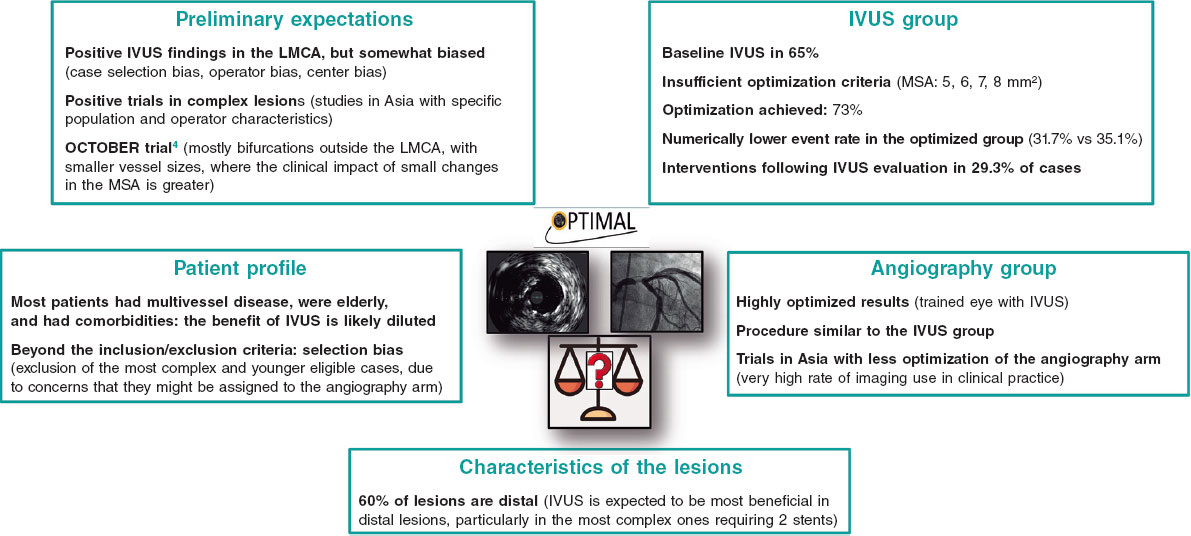
A: OPTIMAL1 was a randomized, multicenter, open-label trial comparing angiography-guided with intravascular ultrasound (IVUS)-guided percutaneous coronary intervention in patients with left main coronary artery disease. The trial enrolled a total of 806 patients and was conducted at European centers with extensive experience in IVUS use. The primary endpoint was a patient-oriented composite endpoint of stroke, acute myocardial infarction, any revascularization, and all-cause mortality. The study population included a high proportion of patients with multivessel disease and a mean SYNTAX score of 30 in both groups. These are two factors that may help explain the lack of difference in the primary endpoint between the angiography and IVUS arms. In a high-risk population with multivessel disease, it is difficult to isolate the effect of IVUS on the angioplasty of the left main coronary artery without accounting for the impact of the remaining lesions and whether they were treated under imaging guidance. In addition, these highly complex patients had a very high event rate, with all-cause mortality rates of approximately 15%. This raises the question of whether a patient-oriented primary endpoint of all-cause mortality or any revascularization is the most appropriate measure for assessing the effect of IVUS on the angioplasty of the left main coronary artery.
Another relevant issue is that the optimization criteria used were based on older studies conducted in Asian populations and may not be fully applicable to European patients. In fact, a recently published consensus document has updated recommendations on the minimum stent area criteria for the angioplasty of the left main coronary artery.3
IVUS-CHIP2 was a randomized, multicenter, open-label trial comparing angiography-guided with IVUS-guided percutaneous coronary intervention in patients undergoing complex procedures, including severe calcification, ostial lesions, bifurcation lesions ≥ 2.5 mm, left main coronary artery disease, chronic total coronary occlusion, restenosis, lesions > 28 mm, and percutaneous coronary intervention requiring mechanical circulatory support. The study included a total of 2020 patients and was conducted at European high-volume centers with high IVUS utilization. The primary endpoint was target vessel failure defined as a composite of cardiac death, target vessel myocardial infarction, and clinically driven target vessel revascularization. Long lesions were the most common complex lesion type, accounting for almost 65% of cases. Although the authors reported severe calcification in approximately 40% of lesions, this contrasts with the relatively low use of advanced plaque-modification techniques, such as rotational atherectomy, which was used in only 6%-7% of cases. Again, no significant differences in the primary endpoint were observed between the IVUS-guided and angiography-guided groups. However, the IVUS arm showed a significant reduction in stent thrombosis. An important feature of IVUS-CHIP was the low proportion of patients in whom adequate optimization according to the predefined criteria was achieved, only 48%. The main reason for failure to meet the optimization criteria was residual plaque burden > 50% at the distal edge. This may have reflected the lack of preintervention imaging to select appropriate landing zones, suboptimal interpretation of imaging findings by operators, or the inclusion of diffuse disease, particularly since long lesions were the most frequently represented lesion type.
Several features common to both trials may help explain the lack of difference between the imaging and angiography arms. The first is the limited impact of imaging on operator behavior. Both studies reported very similar stent sizes, postdilation rates, and use of plaque-modification techniques. This suggests that imaging did not substantially alter how operators behaved. In fact, in a large proportion of cases, imaging was performed only after stenting, which markedly reduced its ability to guide the procedure. Intracoronary imaging is not beneficial simply because the catheter is introduced; the operator must interpret the findings and adapt the procedure accordingly. If the procedure was essentially the same in both groups, it is unsurprising that clinical outcomes were similar as well. Another striking finding in both trials is that IVUS use increased procedural time by more than 30%. This is notable because interpretation of the basic parameters used to optimize percutaneous coronary intervention should not substantially prolong procedural time at experienced centers, particularly when imaging was performed only after stenting in a considerable proportion of patients. External validity is another important consideration. These results were obtained at centers with extensive experience in intracoronary imaging, where operators are likely to have already incorporated part of that knowledge into procedures performed under angiographic guidance alone. It is therefore reasonable to ask whether operators with less experience in intracoronary imaging would perform angiography-guided procedures in the same way and achieve similar results.
Q: In light of these findings, what is your current recommendation on the use of intravascular imaging in percutaneous coronary intervention?
A: In my view, these results do not change the indications for intracoronary imaging. There is substantial prior evidence showing that intracoronary imaging can improve outcomes in complex lesions and left main coronary artery disease when the findings are used to guide treatment. Appropriate lesion selection for imaging and accurate interpretation of the findings are essential if the imaging modality is to deliver its full clinical benefit.
Q: Is the available clinical evidence comparable for IVUS and optical coherence tomography (OCT)?
A: There is evidence supporting both imaging modalities. In my opinion, however, the choice between the 2 should depend on lesion type, operator experience with each modality, and patient- specific factors.
Q: In which settings do you consider one imaging modality preferable to the other, and what are your own preferences in clinical practice?
A: Overall, OCT is the preferred imaging modality when high resolution and detailed characterization are required, for example to assess the pattern of calcification or identify the type of intrastent tissue. In my practice, OCT is particularly useful for evaluating the mechanisms of stent failure, including in-stent restenosis and thrombosis, characterizing the underlying substrate and guiding percutaneous coronary intervention in patients with acute coronary syndrome. By contrast, IVUS remains my preferred imaging modality for ostial lesions, both in left main and right coronary arteries, as well as in situations where contrast use should be minimized or injections avoided, such as chronic total coronary occlusion. Both imaging modalities are useful for long lesions, calcified lesions, and bifurcations.
FUNDING
None declared.
STATEMENT ON THE USE OF ARTIFICIAL INTELLIGENCE
Artificial intelligence was not used in the preparation of this article.
CONFLICTS OF INTEREST
The author reports having received honoraria as a consultant and speaker from Abbott, Boston Scientific, Shockwave, Philips, Cordis, and SMT.
REFERENCES
1. Testa L, De la Torre Hernandez JM, De Maria GL, et al. IVUS-Guided versus Angiography-Guided PCI in Unprotected Left Main Coronary Disease. N Engl J Med. 2026;394:2189-2199.
2. Diletti R, Daemen J, Faurie B, et al. Intravascular Ultrasound-Guided or Angiography-Guided Complex High-Risk PCI. N Engl J Med. 2026;394:2200-2211.
3. Johnson TW, Gonzalo N, de la Torre Hernandez JM, et al. Intracoronary imaging for left main percutaneous coronary intervention: a clinical consensus statement of the European Association of Percutaneous Cardiovascular Interventions (EAPCI) of the ESC and the European Bifurcation Club (EBC). Eur Heart J. 2026;47:5247-5265.