
To the Editor,
Post-percutaneous coronary intervention (PCI) physiological assessment provides important prognostic information, with lower postprocedural instantaneous wave-free ratio (iFR) values associated with adverse clinical outcomes.1 Although virtual prediction of post-PCI iFR is increasingly used for procedural planning, the temporal behavior of iFR immediately after stent deployment remains poorly defined.2 We therefore investigated the kinetics of post-PCI iFR, the timing of physiological stabilization, the agreement between virtual and observed post-PCI iFR, and the relationship between balloon ischemic time and postprocedural iFR recovery. The study was approved by the local ethics committee.
We prospectively studied a total of 107 consecutive patients undergoing iFR-guided PCI for intermediate de novo noncalcified coronary lesions with a pre-PCI iFR ≤ 0.89. The mean age was 65.1 ± 12.9 years, and 63.0% of patients were men. Hypertension (66.7%) and dyslipidemia (51.9%) were the most prevalent cardiovascular risk factors, followed by diabetes mellitus (24.1%) and obesity (21.3%). Previous myocardial infarction and PCI were present in 25.9% and 35.2% of patients, respectively. Most procedures were performed for stable angina (75.9%), whereas 23.1% were performed for non-ST-segment elevation myocardial infarction. The treated vessels were the left anterior descending artery (48.6%), left circumflex (31.8%), and right coronary arteries (19.6%). Lesion preparation was performed with semicompliant or noncompliant balloons in all cases.
Coronary iFR assessment and angiographic co-registration were performed using the SyncVision software (version 4.1.0.5; Philips Volcano, Belgium). The iFR was measured using a pressure guidewire equipped with a distal pressure sensor to simultaneously record aortic and distal coronary pressures during the wave-free period of diastole, when coronary resistance is naturally minimized and the pressure-flow relationship is stable. The distal iFR value was calculated as the mean of 5 consecutive cardiac cycles. Subsequently, iFR pullback with angiographic co-registration was performed by slowly withdrawing the pressure wire under fluoroscopic guidance toward the guiding catheter while continuously recording pressure measurements along the vessel. The software automatically co-registered the physiological pullback data with the corresponding angiographic images, enabling visualization of the location and magnitude of pressure gradients throughout the coronary artery. This physiological map facilitates virtual PCI planning by allowing the operator to simulate stent placement and estimate the predicted distal post-PCI iFR. Lesions with an iFR ≤ 0.89 were considered hemodynamically significant, and PCI was recommended in these patients.3 After final angiography confirmed a good angiographic result, the guiding catheter was flushed with saline to prevent damping of the pressure waveform. After stabilization of the pressure waveform, serial iFR measurements were obtained every minute for 10 minutes after the final balloon inflation while maintaining the distal wire in a fixed position. Balloon ischemic time was defined as the cumulative duration of all balloon inflations, expressed in seconds.
Mean pre-PCI iFR was 0.743 ± 0.125. Immediately after PCI, iFR was substantially lower than the final stabilized value, increasing from 0.899 ± 0.032 at 1 minute to 0.933 ± 0.023 at 10 minutes; P < .001. The greatest increases occurred within the first 3 minutes, with smaller but still significant increases through minute 5. Thereafter, consecutive measurements no longer differed significantly, indicating physiological stabilization approximately 5 minutes after stent deployment (figure 1). Overall, 89.7% of patients showed an increase in iFR > 0.01 between the first post-PCI measurement and the peak value, with a mean increase of 0.038 ± 0.022.
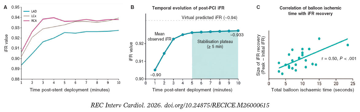
Figure 1. Temporal evolution of post-PCI iFR and its relationship with balloon ischemic time. A: temporal changes in mean post-PCI iFR according to the treated coronary artery. Left anterior descending coronary artery (LAD) lesions showed the lowest initial post-PCI iFR values and the greatest delayed physiological recovery, whereas the left circumflex (LCx) and right coronary artery (RCA) lesions reached higher iFR values and an earlier plateau. B: overall temporal evolution of mean post-PCI iFR after stent implantation. Mean iFR increased rapidly within the first 3 to 5 minutes after PCI, followed by a stabilization plateau from approximately 5 minutes onward, approaching the virtual predicted post-PCI iFR (~0.94). C: correlation between total balloon ischemic time and the magnitude of iFR recovery, defined as the difference between peak and initial post-PCI iFR values. Longer cumulative balloon inflation time was associated with greater delayed physiological recovery (Pearson r = 0.50; P < .001), suggesting that transient ischemia during PCI contributes to reversible postprocedural depression of coronary physiology. iFR, instantaneous wave-free ratio; PCI, percutaneous coronary intervention; SD, standard deviation.
Overall physiological values differed according to coronary territory, with left circumflex artery (LCx) and right coronary artery (RCA) lesions showing higher post-PCI iFR values than left anterior descending coronary artery (LAD) lesions. A significant time-by-vessel interaction (P = .003) indicated that the temporal recovery profile differed among vessels, with RCA lesions reaching physiological stabilization earlier, whereas LAD lesions showed a more gradual improvement.
The virtual predicted post-PCI iFR was 0.944 ± 0.022 modestly exceeding the highest observed post-PCI iFR of 0.937 ± 0.022, with a mean difference of 0.007 ± 0.013; P < .001. However, the virtual predicted value correlated strongly with the observed value (r = 0.84; P < .001), supporting its usefulness as an estimate of achievable physiological improvement.
Balloon ischemic time correlated positively with the magnitude of post-PCI iFR recovery (Pearson r = 0.50; P < .001). Linear regression showed an approximate increase of 0.020 iFR units for every additional 10 seconds of cumulative balloon inflation. Patients with longer balloon inflation times showed greater recovery from the early post-PCI measurements, suggesting a greater degree of transient physiological suppression immediately after PCI.
Transient myocardial ischemia induced by coronary balloon inflation is known to produce reactive microvascular hyperemia that is more intense and prolonged than that achieved with conventional adenosine administration.4 Consequently, true resting physiological conditions may not be present immediately after PCI, resulting in artificially low iFR values. Our findings demonstrate that post-PCI iFR is a dynamic parameter and may substantially underestimate the final physiological result when measured immediately after stent implantation. Physiological stabilization occurred approximately 5 minutes after PCI, suggesting that measurements obtained earlier may not accurately reflect the final postprocedural physiological state. Furthermore, the association between balloon ischemic time and iFR recovery supports the hypothesis that transient balloon-induced microvascular hyperemia contributes to the early underestimation of physiological improvement when resting indices are measured immediately after PCI.
This study has several limitations, including its single-center design and the absence of long-term clinical outcome data. Furthermore, the 10-minute observation period may not have been sufficient to fully characterize the temporal evolution of post-PCI iFR because physiological recovery may continue beyond the study observation period. Nevertheless, these findings suggest that post-PCI physiological assessment should ideally be deferred for at least 3 to 5 minutes after stent deployment, particularly in procedures involving prolonged balloon inflation times.
FUNDING
No funding was received for this study.
ETHICAL CONSIDERATIONS
This study was approved by the appropriate ethics committee. Informed consent was obtained from all participants. Potential sex- and gender-related biases were taken into consideration in the preparation of this work.
STATEMENT ON THE USE OF ARTIFICIAL INTELLIGENCE
Large language models were used to improve the readability and language of the manuscript.
AUTHORS’ CONTRIBUTIONS
D. Faria: study design, patient recruitment and data collection, statistical analysis, and manuscript drafting. M. Santos: patient recruitment and data collection. S. Bravo Baptista: manuscript review. H. Vinhas: study supervision and manuscript review.
CONFLICTS OF INTEREST
D. Faria declared no conflicts of interest whatsoever. M. Santos declared no conflicts of interest whatsoever. S. Bravo Baptista has received research grants from Philips, Abbott, and Xeltis; honoraria and speaker fees from Philips; and consulting and advisory board fees from Abbott and Philips. H. Vinhas declared no conflicts of interest whatsoever.
REFERENCES
1. Patel MR, Jeremias A, Maehara A, et al. 1-Year Outcomes of Blinded Physiological Assessment of Residual Ischemia After Successful PCI: DEFINE PCI Trial. JACC Cardiovasc Interv. 2022;15:52-61.
2. Kikuta Y, Cook CM, Sharp ASP, et al. Pre-Angioplasty Instantaneous Wave-Free Ratio Pullback Predicts Hemodynamic Outcome In Humans With Coronary Artery Disease: Primary Results of the International Multicenter iFR GRADIENT Registry. JACC Cardiovasc Interv. 2018;11:757-767.
3. Vasiljevs D, Kakurina N, Pontaga N, et al. Culprit versus Complete Revascularization during the Initial Intervention in Patients with Acute Coronary Syndrome Using a Virtual Treatment Planning Tool: Results of a Single-Center Pilot Study. Medicina (Kaunas). 2023;59:270.
4. Stoller M, Seiler C. Reactive myocardial hyperaemia for functional assessment of coronary stenosis severity. EuroIntervention. 2017;13:e201-e209.