
To the Editor,
In patients with severe symptomatic mitral regurgitation (MR) at high surgical risk, mitral transcatheter edge-to-edge repair (M-TEER) has become the standard of care. However, up to 50% of these patients are deemed ineligible because of anatomical constraints, including severe calcification or insufficient leaflet length.1 Transcatheter mitral valve replacement (TMVR) has emerged as a promising alternative capable of virtually abolishing MR regardless of its etiology. Early TMVR systems predominantly required a transapical approach, which was associated with substantial morbidity, and carried a high risk of left ventricular outflow tract (LVOT) obstruction. The Edwards SAPIEN M3 system (Edwards Lifesciences, United States) represents a major advance in TMVR. It is a fully transseptal, balloon-expandable TMVR system that uses a nitinol dock encircling the chordae tendineae to create a landing zone in which a 29-mm SAPIEN valve is deployed.2 This design preserves the native subvalvular apparatus, minimizes the risk of LVOT obstruction, and improves prosthesis anchoring compared with other commercially available transcatheter mitral prostheses, particularly in patients with native mitral valve disease. Available data remain scarce and are limited to the Early Feasibility Study3 and the recently published pivotal ENCIRCLE trial.4 We describe our early clinical and echocardiographic experience in high-risk patients undergoing TMVR with the SAPIEN M3 system.
We conducted a retrospective analysis of 6 consecutive patients who underwent TMVR with the SAPIEN M3 system between July 2025 and March 2026 (figure 1). Seven additional patients did not meet the inclusion criteria (53.8%; 7 of 13 screened), with a high risk of LVOT obstruction being the leading reason for exclusion. This retrospective study was approved by the Ethics Committee of Hospital Universitario Ramón y Cajal (Madrid, Spain). All patients were informed about procedural risks and expected benefits and provided written informed consent prior to the intervention. All candidates underwent computed tomography (CT)–based imaging screening to assess anatomical suitability and estimate the neo-LVOT area. Postdischarge imaging follow-up included a CT sca at 30-60 days and serial transthoracic echocardiography. The antithrombotic protocol consisted of continuing vitamin K antagonists in patients already receiving this therapy. In patients previously receiving non–vitamin K antagonist oral anticoagulants, apixaban was prescribed at discharge because of its twice-daily dosing regimen. Suspected or confirmed device thrombosis prompted a switch to a vitamin K antagonist, with low-dose oral acetylsalicylic acid (100 mg/d) added if thrombosis persisted despite a therapeutic international normalized ratio (INR).
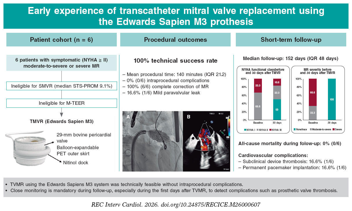
Figure 1. Baseline characteristics, procedural outcomes, and short-term follow-up findings after TMVR with the Edwards SAPIEN M3 system. A: balloon inflation after adequate positioning of the device within the previously implanted dock. B: intraprocedural TEE color Doppler image obtained after valve implantation, showing adequate device position and no residual MR. IQR, interquartile range; MR, mitral regurgitation; M-TEER, mitral transcatheter edge-to-edge repair; NYHA, New York Heart Association; PET, polyethylene terephthalate; SMVR, surgical mitral valve replacement; STS-PROM, Society of Thoracic Surgeons Predicted Risk of Mortality; TEE, transesophageal echocardiography; TMVR, transcatheter mitral valve replacement.
The median age was 76 years (IQR, 4.5 years), and 33.3% of the patients were women. Comorbidities included atrial fibrillation in 100% of patients and previous coronary artery bypass grafting in 33.3%. The estimated surgical risk was prohibitively high, with a median 30-day Society of Thoracic Surgeons Predicted Risk of Mortality (STS-PROM) score of 9.1%. The etiology was ischemic secondary MR in 3 patients and degenerative primary MR in the remaining 3. Importantly, the entire cohort was considered unsuitable for M-TEER: 5 patients because of primary anatomical constraints, such as leaflet thickening or insufficient posterior leaflet length, and 1 because of a previous unsuccessful M-TEER procedure. Baseline echocardiography showed a preserved median left ventricular ejection fraction of 53% (IQR, 18.2%). All patients also had concomitant tricuspid regurgitation at baseline, which was mild in 50% and moderate in 50%.
Under general anesthesia and transesophageal echocardiographic and fluoroscopic guidance, the nitinol dock was introduced via right femoral venous access using a 23-Fr system and successfully deployed around the subvalvular apparatus. The 29-mm SAPIEN valve was subsequently expanded within the dock. Technical success was achieved in all patients, with a median procedural duration of 140.5 minutes. Intraprocedural hemodynamic findings were highly favorable. Transesophageal echocardiography confirmed an immediate reduction in MR to none or trace in all patients, without intraprocedural complications or evidence of LVOT obstruction.
Clinical and echocardiographic follow-up was performed over a median of 152 days (IQR, 48 days). The abolition of MR resulted in substantial clinical improvement. At 30 days, 50% of the patients had a clinically meaningful improvement of at least 1 New York Heart Association (NYHA) functional class (table 1). Echocardiographic assessment confirmed the sustained effectiveness of the intervention, with MR remaining absent or trace in 100% of the cohort. A mild septal paravalvular leak was detected in 1 patient (16.6%). No cases of hemolysis occurred, and closure of the iatrogenic atrial septal defect was not required in any of the 6 patients. The median transmitral gradient was favorable at 3.9 mm Hg (IQR, 1.3 mm Hg).
Table 1. Comparison of baseline and 30-day clinical, echocardiographic, and CT-derived data
| Variable | Baseline (n = 6) | 30-day follow-up (n = 6) |
|---|---|---|
| NYHA functional class | ||
| I | — | 3 (50.0) |
| II | 4 (66.7) | 2 (33.3) |
| III | 2 (33.3) | 1 (16.7) |
| Improvement in NYHA functional class | — | 4 (66.7) |
| MR severity | ||
| None or trace | — | 5 (100) |
| Mild | — | — |
| Moderate to severe | 2 (33.3) | — |
| Severe | 4 (66.7) | — |
| 2D MR EROA, cm2 | 0.34 (0.20) | — |
| 2D MR vena contracta, mm | 6.3 (1.4) | — |
| MR PISA radius, mm | 9.9 (1.2) | — |
| MR regurgitant volume, mL | 47.9 (23.5) | — |
| Mean transmitral pressure gradient, mm Hg | 2.7 (0.7) | 4.7 (1.4) |
| Concomitant valvular heart disease | ||
| Mitral stenosis | 0 (0) | — |
| Aortic stenosis | 2 (33.3)a | — |
| Aortic regurgitation | 1 (16.7)b | — |
| Tricuspid regurgitation | 6 (100)c | 6 (100) |
| Severe or greater tricuspid regurgitation | 0 (0) | 2 (33.3) |
| Maximum TR velocity, m/s | 3.1 (0.6) | 3.18 (0.10) |
| Mean LVOT gradient, mm Hg | 1.5 (0.4) | 2.6 (2.9) |
| Baseline CT data | ||
| Anteroposterior diameter, mm | 35.9 (3.3) | — |
| Commissure-to-commissure diameter, mm | 42.0 (3.0) | — |
| Mitral annular perimeter, mm | 121.3 (22.2) | — |
| MVA, cm2 | 12.0 (2.7) | — |
| Aortomitral angle, degrees | 129.0 (5.3) | — |
| MAC, Guerrero score | 1.5 (3.2) | — |
| Predicted neo-LVOT area, mm2 | 456.5 (158.7) | — |
|
Qualitative variables are expressed as frequency (percentage) and quantitative variables as median (interquartile range). EROA, effective regurgitant orifice area; LA; left atrial; LVEDV; left ventricular end-diastolic volume; LVEF; left ventricular ejection fraction; LVOT; left ventricular outflow tract; MAC, mitral annular calcification; MR; mitral regurgitation; MVA, mitral valve area; NYHA; New York Heart Association; PVL; paravalvular leak; RV; right ventricular; TAPSE; tricuspid annular plane systolic excursion; TR; tricuspid regurgitation. a Mild aortic stenosis. b Mild aortic regurgitation. c 3 patients had moderate TR and 3 patients had mild TR. |
||
The median left ventricular ejection fraction increased slightly from 53% to 55%, accompanied by a reduction in left ventricular end- diastolic volume from 94.5 mL to 85 mL. However, assessment of the right-sided cardiac chambers revealed a heterogeneous response after the abrupt correction of volume overload. Although overall right ventricular dimensions decreased slightly, with the basal diameter decreasing from 41.5 mm to 41 mm, and longitudinal systolic function improved marginally, with tricuspid annular plane systolic excursion increasing from 18 mm to 20 mm, concomitant tricuspid regurgitation progressed to severe in 2 patients (33.3%). Both patients were managed with optimized medical therapy and remain under close outpatient surveillance, with transcatheter tricuspid intervention reserved for cases in which it becomes necessary.
Regarding safety outcomes, patient #1, who had preexisting right bundle branch block, developed complete atrioventricular block on postoperative day 3, requiring permanent pacemaker implantation. Before discharge, the heart team decided to switch anticoagulation from apixaban to acenocoumarol because of a suspected increased risk of valve thrombosis, based on a mean gradient of 5.5 mm Hg across the M3 prosthesis. Despite adequate INR values after discharge (INR, 3.0-3.3), transesophageal echocardiography performed on day 14 showed prosthetic leaflet thickening and subtle restriction of the anteriorly positioned leaflet. After confirmation by CT scan, low-dose oral acetylsalicylic acid (100 mg/d) was added. The patient experienced no embolic events or worsening heart failure symptoms. Follow-up echocardiography and CT performed 4 and 6 months after TMVR, respectively, showed partial resolution of leaflet thickening and improved leaflet mobility.
Our initial experience is consistent with the early efficacy findings of the pivotal ENCIRCLE trial,4 with a technical success rate of 100%, confirming the feasibility of the transseptal dock-and-valve approach. The ability to completely abolish MR highlights the potential advantage of TMVR over M-TEER in achieving immediate hemodynamic correction, especially in complex anatomies traditionally considered unsuitable for edge-to-edge repair. However, the safety profile observed in our real-world cohort highlights important aspects of post-TMVR management. The incidence of conduction disturbances is a known challenge. Although the ENCIRCLE trial reported a low permanent pacemaker implantation rate of 2.6%, the rate in our cohort was 16.6%. This difference likely reflects the small sample size, the presence of baseline conduction disease, and mechanical stress exerted by the nitinol frame on periannular conduction tissue, underscoring the need for close electrocardiographic monitoring.
Another important finding was the early occurrence of subclinical device thrombosis, which has been reported in approximately 6.7% to 12.3% of patients after TMVR.5 Post-TMVR antithrombotic management remains controversial. The large prosthetic surface area, low left atrial flow, and presence of atrial fibrillation in all patients create a highly thrombogenic environment. Detection of restricted leaflet motion on day 14 highlights the need for rigorous anticoagulation regimens and a low threshold for follow-up imaging.
Finally, the dynamic interaction between correction of left-sided valve disease and the progression of right-sided valve disease is highly complex. The paradoxical worsening of severe tricuspid regurgitation to severe grades, reported in approximately 16% of patients after transcatheter mitral interventions in recent studies,6 occurred in 33.3% of our cohort despite improvements in left-sided hemodynamics and right ventricular longitudinal function, suggesting a multifactorial etiology. Proposed mechanisms include an abrupt increase in cardiac output, interventricular septal shift, and mechanical annular distortion, underscoring the need for comprehensive right heart assessment.
In conclusion, transseptal TMVR with the SAPIEN M3 system is a feasible, safe, and hemodynamically effective alternative for patients with high-risk MR who are ineligible for M-TEER. However, the postprocedural course requires close surveillance because of the risks of conduction disturbances, subclinical device thrombosis, and unpredictable progression of concomitant tricuspid regurgitation. Further studies are needed to clarify these findings.
FUNDING
No funding was received for this study.
ETHICAL CONSIDERATIONS
This retrospective study was approved by the Ethics Committee of Hospital Universitario Ramón y Cajal (Madrid, Spain). All patients gave their written informed consent before the procedure. Patient data were collected and reported in full compliance with the CARE case report guidelines and the ethical principles of the Declaration of Helsinki. Potential sex- and gender-related biases were considered in accordance with the SAGER guidelines. The authors accept the responsibilities established by the International Committee of Medical Journal Editors (ICMJE).
STATEMENT ON THE USE OF ARTIFICIAL INTELLIGENCE
The authors declare that artificial intelligence was not used in the preparation of this manuscript, in accordance with the recommendations of the International Committee of Medical Journal Editors (ICMJE).
AUTHORS’ CONTRIBUTIONS
Á. Sánchez-Recalde was responsible for the conceptualization and design of the study, supervision of the study process, and approval of the final version of the manuscript. A. Larrea Iñarra contributed to data collection and statistical analysis and drafted the manuscript. L. Salido Tahoces contributed to the statistical analysis and clinical follow-up of the patients. A. González Gómez was responsible for the clinical assessment and postprocedural follow-up of the patients. C. Fernández-Golfín Lobán and J.L. Zamorano Gómez supervised the study process and reviewed and approved the final version of the manuscript.
CONFLICTS OF INTEREST
The authors declare no conflicts of interest related to the publication of this article.
REFERENCES
1. Iung B, Delgado V, Rosenhek R, et al. Contemporary presentation and management of valvular heart disease:The EUrobservational research programme valvular heart disease II survey. Circulation. 2019;140:1156-1169.
2. Tartaglia F, Antonelli G, Gitto M, et al. TMVR for the Treatment of Mitral Regurgitation:A State-of-the-Art Review. Circ Cardiovasc Interv. 2025;18:e015298.
3. Del Val D, Ferreira-Neto AN, Wintzer-Wehekind J, et al. Early experience with transcatheter mitral valve replacement:A systematic review. J Am Heart Assoc. 2019;8:e013332.
4. Guerrero ME, Daniels DV, Makkar RR, et al. Percutaneous transcatheter valve replacement in individuals with mitral regurgitation unsuitable for surgery or transcatheter edge-to-edge repair:a prospective, multicountry, single-arm trial. Lancet. 2025;406:2541-2550.
5. Kikoïne J, Urena M, Chong-Nguyen C, et al. Predictors and clinical impact of thrombosis after transcatheter mitral valve implantation using balloon-expandable bioprostheses. EuroIntervention. 2021;16:1455-1462.
6. Adamo M, Pagnesi M, Ghizzoni G, et al. Evolution of tricuspid regurgitation after transcatheter edge-to-edge mitral valve repair for secondary mitral regurgitation and its impact on mortality. Eur J Heart Fail. 2022;24:2175-2184.