
This is the case of a 67-year-old man with signs of right heart failure due to an atrial septal defect (ASD) with right ventricular dilation, significant left-to-right shunt (Qp/Qs 1.5), and no significant pre-capillary pulmonary hypertension (mean pulmonary artery pressure, 26 mmHg; pulmonary vascular resistance, 2UW). Three-dimensional transesophageal echocardiography (3DTEE) revealed the presence of a cor triatriatum sinister (CTS) with a membrane extending from the upper-posterior end of the fossa ovalis to the pulmonary ridge, with multiple fenestrations, the largest measuring 24 mm (figure 1A). A 22 mm × 11 mm ostium secundum ASD was confirmed with the posterior rim partially covered by the membrane (figure 1B,C). Since the patient was technically suitable and had increased surgical risk due to cirrhosis, a percutaneous approach was undertaken. The procedure was performed under 3DTEE and fluoroscopy guidance, a 0.035-in guidewire and multipurpose catheter crossed the ASD, balloon sizing confirmed a 20 mm defect, and an 24 mm Amplatzer septal occluder (Abbott Structural Heart, United States) was successfully implanted (figure 1D,F). TEE confirmed accurate device placement, with no residual shunt and no left atrial flow obstruction. Informed consent was obtained.
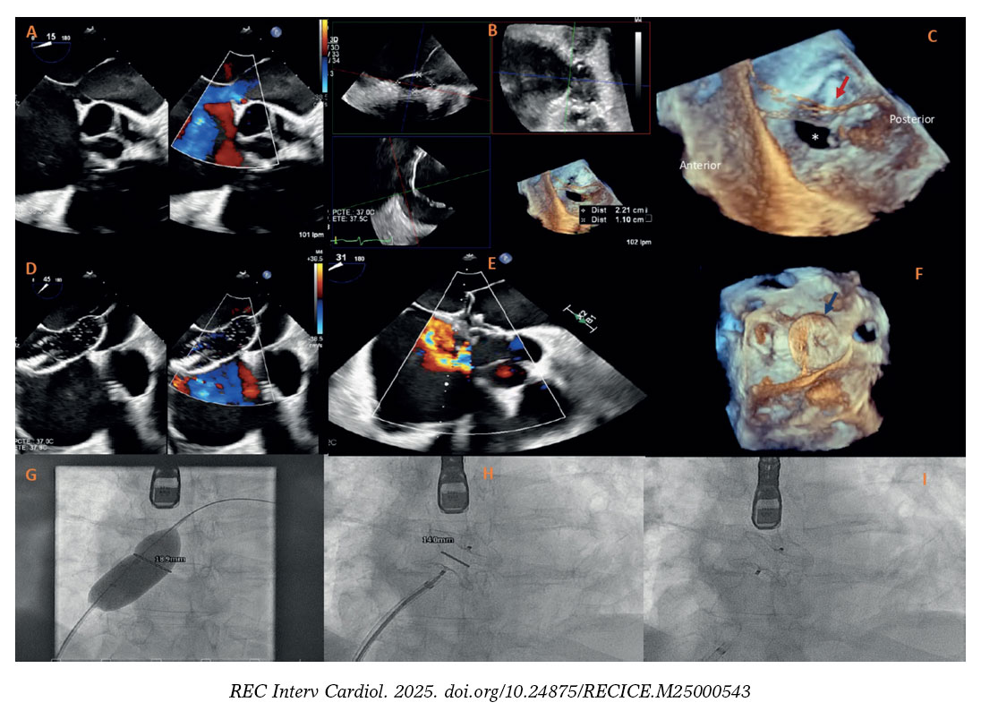
Figure 1.
CTS is a rare cardiac malformation that can coexist with ostium secundum ASD in up to 33% of patients. Although transcatheter closure is preferred in suitable anatomies, the presence of CTS can complicate the device deployment due to inadequate margin definition, with increased risk of device instability and embolization. In this scenario, ASD closure was particularly challenging as the posterior rim was in direct continuity with the atrial membrane, raising concerns about proper device apposition. This case highlights the importance of advanced imaging, such as 3DTEE, in complex anatomies by allowing precise procedural planning and real-time guidance to ensure a successful intervention.
FUNDING
None declared.
ETHICAL CONSIDERATIONS
The patient gave his prior written informed consent for the procedure and for the publication of this case report, including all associated images. The SAGER guidelines have been followed with respect to possible sex/gender bias.
STATEMENT ON THE USE OF ARTIFICIAL INTELLIGENCE
Artificial intelligence, including language models, such as ChatGPT by OpenAI were used solely to support language editing of the manuscript. The scientific content, analysis, and interpretation were entirely produced by the authors.
AUTHORS’ CONTRIBUTIONS
A. Abrantes was responsible for the study conception, literature review, manuscript writing, image processing, and figure preparation. E. Colón contributed to manuscript revision, image processing, and figure preparation. E. Pozo Osinalde performed the transesophageal echocardiography, acquired the echocardiographic images, and contributed to manuscript revision and figure preparation. D. García-Arribas was responsible for the patient’s clinical follow-up and manuscript revision. L. Nombela-Franco and Pablo Salinas performed the interventional procedure and contributed to manuscript revision. All authors revised and approved the final version.
CONFLICTS OF INTEREST
None declared.


