
To the Editor,
Radial access has become the preferred route for diagnostic and interventional coronary procedures because it is associated with lower rates of vascular and bleeding complications compared with femoral access. However, within the radial approach, the choice between right radial access (RRA) and left radial access (LRA) still depends largely on operator experience, cath lab organization, and patient characteristics.
This issue may be particularly relevant in elderly patients.1 Aortic elongation, vascular calcification, and tortuosity of the right brachiocephalic trunk can hinder catheter advancement and manipulation through the RRA.2 Consequently, some observational studies have suggested that LRA may reduce fluoroscopy times, contrast volume, and the need for access crossover. However, the available evidence is heterogeneous, and randomized studies specifically involving elderly patients remain scarce.
To compare the 2 access routes in this population, we conducted a single-center, prospective, randomized study of patients older than 75 years undergoing diagnostic coronary angiography between May 2019 and January 2024. Both outpatients and inpatients referred electively were eligible. Patients were excluded if they required emergency procedures, had hemodynamic instability, a history of coronary artery bypass grafting, no palpable radial pulse, an arteriovenous fistula in either arm, known vascular anatomy favoring a specific access route, or were unable to provide informed consent. The study was conducted in full compliance with the principles outlined in the Declaration of Helsinki. The protocol was approved by the ethics committee of Hospital 12 de Octubre (Madrid, Spain) and all participants gave their written informed consent before enrollment.
Patients were randomized to coronary angiography via the RRA or LRA using a computerized random-number generator. Procedures were performed using the standard Seldinger technique, with 5-Fr or 6-in-5-Fr hydrophilic introducer sheaths selected at the operator’s discretion. We recorded procedure and fluoroscopy times, radiation dose, contrast volume, number of catheters used, crossover to the contralateral access route, periprocedural complications, and patient-perceived pain on a numerical rating scale.
Of the 277 patients randomized, 270 were ultimately analyzed because only 1 coronary artery was selectively engaged in 7 cases. Mean age was 83.8 ± 4.6 years, and 47.9% of the patients were women. A total of 136 patients were assigned to the RRA group and 134 to the LRA group. Baseline characteristics were similar between the 2 groups. Most procedures were performed using a 5-Fr introducer sheaths and conventional diagnostic catheters.
There were no statistically significant differences between the RRA and LRA groups in total procedure time (12.0 ± 7.0 vs 11.3 ± 4.7 minutes; P = .36), time to complete examination of the first coronary artery (7.2 ± 4.0 vs 7.2 ± 4.5 minutes; P = .93), or time to complete examination of the second coronary artery (4.7 ± 4.3 vs 4.1 ± 4.5 minutes; P = .15). Fluoroscopy time was 4.8 ± 3.7 minutes in the RRA group and 4.0 ± 3.2 minutes in the LRA group, although the difference did not reach statistical significance (P = .06).
Furthermore, contrast volume was similar between the 2 approaches (75.6 ± 21.8 mL with RRA vs 74.2 ± 19.3 mL with LRA; P = .59). There were no differences in radiation exposure measured by air kerma (302.2 ± 132.6 vs 317.8 ± 131.7 mGy; P = .35) or dose–area product (22418.6 ± 11740.9 vs 22448.6 ± 11162.5 mGy·cm2; P = .98). The number of catheters used was comparable between the 2 groups (table 1).
Table 1. Variables collected and corresponding P values
| Variable | Overall (n = 270) | Right radial access (n = 136) | Left radial access (n = 134) | P |
|---|---|---|---|---|
| Female sex, % | 47.9 | 47.7 | 48.1 | .96 |
| Age, y | 83.8 ± 4.6 | 83.7 ± 4.7 | 83.9 ± 4.5 | .69 |
| Introducer sheath, % | ||||
| 5-Fr | 73.6 | 76.5 | 70.6 | .32 |
| 6-Fr | 26.4 | 23.5 | 29.4 | |
| Fluoroscopy time, min | 4.4 ± 3.5 | 4.8 ± 3.7 | 4.0 ± 3.2 | .06 |
| Air kerma, mGy | 310.2 ± 132.6 | 302.2 ± 132.6 | 317.8 ± 131.7 | .35 |
| DAP, mGy·cm2 | 22 433.5 ± 11 436.2 | 22 418.6 ± 11 740.9 | 22 448.6 ± 11 162.5 | .98 |
| Left coronary artery time, min | 7.3 ± 5.0 | 7.2 ± 4.0 | 7.2 ± 4.5 | .93 |
| Right coronary artery time, min | 4.4 ± 3.6 | 4.7 ± 4.3 | 4.1 ± 4.5 | .15 |
| Total procedure time, min | 11.6 ± 6.0 | 12.0 ± 7.0 | 11.3 ± 4.7 | .36 |
| Contrast volume, mL | 75.9 ± 20.5 | 75.6 ± 21.8 | 74.2 ± 19.3 | .59 |
| No. of catheters used for the left coronary artery | 1 [1-1] | 1 [1-1] | 1 [1-1] | .94 |
| No. of catheters used for the right coronary artery | 1 [1-1] | 1 [1-1] | 1 [1-1] | .57 |
| Total No. of catheters used | 2 [2-2] | 2 [2-2] | 2 [2-2] | .93 |
| Pain score (1-10) | 2 [1-3] | 2 [1-4] | 2 [1-3] | .73 |
| Crossover to the contralateral access route, % | 4.0 | 5.6 | 2.2 | .15 |
|
DAP, dose-area product. Data are expressed as no. (%), mean ± standard deviation, or median [interquartile range]. |
||||
Crossover to the contralateral access route was required in 5.6% of patients assigned to RRA and 2.2% of those assigned to LRA, with no statistically significant difference (P = .15). In the RRA group, crossover was mainly prompted by difficulty advancing the catheter because of subclavian or right brachiocephalic trunk tortuosity.
There were no major periprocedural complications. One patient developed a forearm hematoma after LRA puncture, with a favorable clinical course, and 4 episodes of radial artery spasm were recorded: 3 with RRA and 1 with LRA. Patient-perceived pain was low and similar in the 2 groups.
The main finding of this study is that, in an elderly population randomized to RRA or LRA for diagnostic coronary angiography, LRA did not demonstrate significant superiority over RRA in the procedural variables analyzed. Specifically, there were no significant differences in total procedure time, coronary cannulation time, contrast volume, radiation exposure, number of catheters used, complications, or patient-perceived pain.
Our findings are consistent with part of the previously published evidence. In the TALENT study, Sciahbasi et al.3 reported shorter fluoroscopy times with LRA, particularly in older patients and when procedures were performed by less experienced operators. By contrast, a randomized study specifically involving octogenarian patients conducted by Freixa et al.4 found no significant differences in most procedural variables between the 2 access routes. Similarly, several meta-analyses have shown modestly favorable results for LRA in fluoroscopy time and contrast volume, although the absolute differences were small and of uncertain clinical relevance.5 Our findings in an exclusively elderly, randomized cohort generally support the view that the potential anatomical advantages of LRA do not necessarily translate into consistent or clinically meaningful procedural benefits.
From a clinical practice perspective, although LRA may be anatomically advantageous in some elderly patients because it avoids passage through the right brachiocephalic trunk, our data do not support its routine use as the preferred access route in this population. Similarly, despite its potentially greater anatomical difficulty in selected patients, RRA achieved similar safety and efficacy outcomes in our study. Both approaches should therefore be considered valid options, and the choice of radial access in elderly patients should be individualized. LRA may be a reasonable option in patients with suspected severe tortuosity of the right brachiocephalic trunk, previous difficulty with RRA, or need to optimize catheter advancement. Conversely, when procedural ergonomics, cath lab layout, or operator experience favor RRA, our findings do not suggest that its use results in worse clinical or procedural outcomes (figure 1).
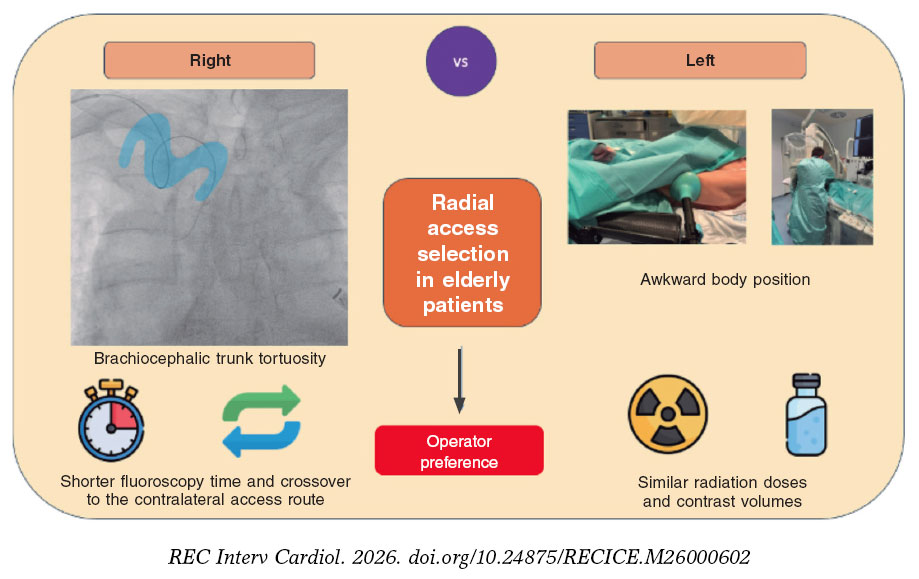
Figure 1. Advantages and disadvantages of left radial access vs right radial access for coronary angiography in elderly patients.
Our study has several limitations. It was conducted at a single-center study and involved operators with different levels of experience. Anatomical variables obtained through systematic vascular imaging were not collected, nor was the radiation dose received by the operator measured directly using personal dosimetry. Moreover, the study was limited to diagnostic coronary angiography, and the findings should not be extrapolated directly to complex interventional procedures. Although the biological sex of participants was recorded and its distribution was balanced between the groups, no specific sex- or gender-based analysis was performed, limiting the assessment of potential differences associated with these variables.
In conclusion, in patients older than 75 years undergoing diagnostic coronary angiography, LRA did not provide statistically significant advantages over RRA in procedure time, radiation exposure, contrast volume, need for additional equipment, complications, or patient-perceived pain. These findings support a pragmatic, individualized strategy to radial access selection in elderly patients, guided by expected vascular anatomy, operator experience, and cath lab organization rather than by the systematic selection of one side over the other.
FUNDING
This study received no funding from any organization.
ETHICAL CONSIDERATIONS
This study was conducted in full compliance with international recommendations for clinical research and was approved by the ethics committee of our institution. Written informed consent was obtained from all patients and archived. Biological sex of participants was recorded, and its distribution was balanced between the groups. However, no specific gender-based analysis was performed, and no information was collected on variables related to gender identity or sociocultural factors.
STATEMENT ON THE USE OF ARTIFICIAL INTELLIGENCE
The authors declare that no artificial intelligence tools were used in the conception, design, data analysis, interpretation, or drafting of this manuscript.
AUTHORS’ CONTRIBUTIONS
C.M. Rivadulla Varela contributed to manuscript drafting and data acquisition. M. Calderón Flores, J. García Tejada, and A. Albarrán González-Trevilla contributed to data acquisition. N.M. Maneiro Melón performed the statistical analysis. M. Velázquez Martín contributed to the conception and design of the manuscript. All authors critically revised the manuscript for important intellectual content and approved the final version. All authors also accept responsibility for all aspects of the work and agree to investigate and resolve any questions concerning the accuracy or integrity of any part of the work.
CONFLICTS OF INTEREST
None declared.
REFERENCES
1. Dehghani P, Mohammad A, Bajaj R, et al. Mechanism and Predictors of Failed Transradial Approach for Percutaneous. Coronary Interventions. JACC Cardiovasc Interv. 2009;2:1057-1064.
2. Kawashima O, Endoh N, Terashima M, et al. Effectiveness of Right or Left Radial Approach for Coronary Angiography. Catheter Cardiovasc Interv. 2004;61:333-337.
3. Sciahbasi A, Romagnoli E, Burzotta F, et al. Transradial approach (left vs right) and procedural times during percutaneous coronary procedures: TALENT study. Am Heart J. 2011;161:172-179.
4. Freixa X, Trilla M, Feldman M, et al. Right versus left transradial approach for coronary catheterization in octogenarian patients. Catheter Cardiovasc Interv. 2012;80:267-272.
5. De Rosa S, Torella D, Caiazzo G, et al. Left radial access for percutaneous coronary procedures: From neglected to performer? A meta-analysis of 14 studies including 7603 procedures. Int J Cardiol. 2014;171:66-72.