
To the Editor,
Drug-coated balloons (DCBs) have revitalized the “leave nothing behind” concept in percutaneous coronary intervention (PCI), enabling the delivery of antiproliferative therapy without a permanent scaffold. Although their efficacy is well established for in-stent restenosis and small-vessel disease, their clinical application has expanded to complex lesion subsets, including chronic total coronary occlusions (CTOs), for which the evidence base remains less mature and vessel wall injury is inherently greater. During CTO PCI, paclitaxel may be inadvertently delivered into the subintimal space or across dissected segments, potentially triggering abnormal remodeling or aneurysm formation. We report cases of focal coronary artery aneurysm (CAA) formation following paclitaxel-coated DCB angioplasty in CTO segments, highlighting a potential mechanistic link between vessel wall injury and antiproliferative drug delivery, and review management considerations for this underrecognized complication.
A 59-year-old man with established ischemic heart disease and previous stenting of the proximal-to-mid right coronary artery (RCA) presented with refractory angina. Coronary angiography revealed a CTO of the RCA distal to the previously implanted stents (figure 1A). Successful antegrade wire escalation was performed using a Gaia Next 2 guidewire (Asahi Intecc, Japan). After balloon angioplasty, intravascular ultrasound (IVUS) demonstrated a segment of subintimal tracking in the mid-to-distal RCA (figure 1B, video S1), although lesion preparation was otherwise adequate. Given the diffusely diseased vessel, extensive previous stenting, the risk associated with the high metallic burden of additional long- segment stenting, and acceptable angiographic luminal gain and flow, a DCB-only strategy was selected after careful consideration. We acknowledge that this represents a clinically contested decision and that stenting with true-lumen scaffolding of the subintimal segment was a legitimate alternative. The mid-to-distal RCA was treated with 2 sequential paclitaxel-coated DCBs (SeQuent Please Neo, B. Braun, Germany; 3.5 mm × 25 mm and 3.0 mm × 25 mm; inflation pressure, 6 atm; inflation time, 60 seconds; overlap, 3-4 mm), achieving Thrombolysis in Myocardial Infarction (TIMI) grade 3 flow with a non-flow-limiting type B dissection according to the National Heart, Lung, and Blood Institute classification (figure 1C). The patient was discharged on dual antiplatelet therapy (aspirin indefinitely and clopidogrel 75 mg daily for 6 months) and remained asymptomatic. Repeat coronary angiography performed at 7 months because of atypical chest pain identified a focal aneurysm in the mid-to-distal RCA, localized to the subintimal segment previously treated with DCB (figure 1D). Invasive physiological assessment confirmed the absence of hemodynamic significance (fractional flow reserve, 0.96) (figure 1E). Repeat intracoronary imaging was not performed because of cost considerations. A conservative strategy was adopted, consisting of continued aspirin monotherapy, optimization of secondary prevention, and planned surveillance with coronary computed tomography angiography (CCTA) at 12 months.
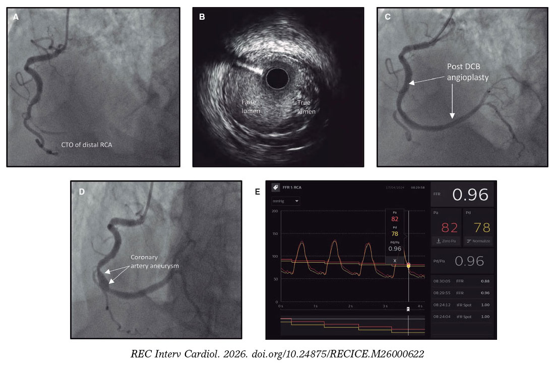
Figure 1. A: baseline angiography of case 1 showing a CTO of the mid-to-distal RCA; B: IVUS of the CTO segment showing extraplaque tracking; C: RCA after paclitaxel-coated DCB angioplasty showing TIMI grade 3 flow; D: RCA at 7-month follow-up showing a focal aneurysm at the subintimal DCB treatment site; E: hemodynamic assessment of the RCA showing nonsignificant indices (fractional flow reserve, 0.96). CTO, chronic total coronary occlusion; DCB, drug-coated balloon; IVUS, intravascular ultrasound; RCA, right coronary artery; TIMI, Thrombolysis in Myocardial Infarction.
In a second case, a 55-year-old man who was an active smoker and had hypertension, dyslipidemia, and previous cerebrovascular disease presented with unstable angina. Coronary angiography demonstrated a CTO of the proximal left anterior descending coronary artery (LAD) (figure 2A), confirmed as a true CTO by blunt stump morphology, bridging collaterals, and a clinical history consistent with an occlusion duration exceeding 3 months. Subacute occlusion was excluded on these grounds. Mild-to-moderate atherosclerotic disease was present in the remaining vessels. Following an initial unsuccessful attempt, repeat CTO intervention was performed 3 months later using parallel-wire and 3-dimensional wiring techniques. IVUS confirmed true-lumen wire position and demonstrated diffuse disease from the ostial to the distal LAD. Lesion preparation using a Wolverine cutting balloon (Boston Scientific, United States) resulted in a localized proximal dissection extending to the media, as confirmed by IVUS (figure 2B, video S2). The dissection was non-flow-limiting, and adequate luminal gain was achieved in the remainder of the vessel. Given the patient’s anatomy—a long LAD CTO with diffuse disease—and the operator’s assessment that extensive stenting from the ostium to the distal LAD carried substantial risk, 3 overlapping paclitaxel-coated DCBs were deployed from the proximal to the distal LAD (Agent, Boston Scientific, United States; 3.5 mm × 30 mm proximally, 3.0 mm × 30 mm in the mid segment, and 2.5 mm × 30 mm distally; inflation pressure, 12 atm; inflation time, 60 seconds; sequential overlaps, 5 mm), achieving an excellent angiographic result with minor residual dissection (figure 2C). The patient was discharged on dual antiplatelet therapy (aspirin indefinitely and ticagrelor 90 mg twice daily for 12 months) and remained asymptomatic. Surveillance angiography at 9 months identified an aneurysmal segment confined precisely to the proximal LAD at the site of the previous dissection (figure 2D). Notably, the remaining DCB-treated LAD segments showed no aneurysmal changes, consistent with vessel wall injury, rather than the DCB drug effect alone, as the primary mechanistic trigger. A conservative strategy was adopted, with further surveillance planned after 12 months.
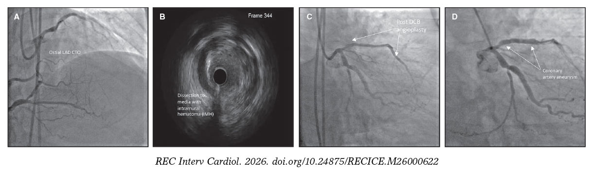
Figure 2. A: baseline angiography of case 2 showing chronic total occlusion of proximal LAD; B: post-cutting balloon IVUS showing medial dissection and intramural hematoma; C: proximal LAD following paclitaxel-coated DCB angioplasty, showing TIMI 3 flow with non-flow limiting dissection; D: proximal LAD at 9-month follow-up showing focal aneurysmal dilation at the DCB treatment site. CTO, chronic total occlusion; DCB, drug-coated balloon; LAD, left anterior descending artery; TIMI, Thrombolysis in Myocardial Infarction.
CAA is defined as focal coronary dilatation exceeding 1.5 times the diameter of the adjacent reference vessel. Although classically associated with atherosclerosis and Kawasaki disease, CAA is a recognized late complication of PCI, occurring in approximately 3.9%-5% of lesions after plain old balloon angioplasty and 0.7%-3.4% after drug-eluting stent implantation. Following treatment with paclitaxel-coated DCBs, aneurysm formation appears uncommon in non-CTO lesions (approximately 0.8%) but may occur more frequently after CTO PCI. Jun et al. reported incidence of 8% following DCB-only CTO intervention, likely reflecting the greater burden of subintimal tracking and vessel wall injury associated with CTO recanalization.1
The underlying mechanism may involve paclitaxel-induced apoptosis of vascular smooth muscle cells. By stabilizing microtubule polymerization, paclitaxel suppresses smooth muscle cell proliferation and migration, processes that are important for neointimal inhibition but also for vessel wall repair.1 Unlike drug-eluting stent implantation, DCB-only treatment leaves the vessel without mechanical support. Previous angiographic and optical coherence tomography (OCT) studies have demonstrated favorable healing of most non-flow-limiting residual dissections after DCB angioplasty, without an increase in adverse clinical outcomes, suggesting that medial dissection alone does not inevitably result in adverse vascular remodeling.2 There are even data suggesting that the absence of OCT-detected medial dissection is associated with target lesion failure after DCB PCI.3 However, these studies largely involved localized dissections in relatively simple de novo lesions. Our cases suggest that extensive vessel wall disruption, including long segments of subintimal tracking or deep extraplaque injury during CTO PCI, may represent a distinct substrate in which impaired reparative responses contribute to aneurysmal remodeling.3,4 This hypothesis remains exploratory and requires further investigation. The localization of aneurysm formation to the dissected segment in case 2, rather than throughout the DCB-treated vessel, further supports the importance of underlying arterial injury, with paclitaxel acting as a contributing rather than an independent factor. Overlapping DCBs, as in case 1, may additionally increase local paclitaxel exposure.
Both aneurysms remained hemodynamically insignificant, and both patients remained asymptomatic throughout follow-up. However, the natural history of DCB-related CAA remains uncertain. It is unclear whether these aneurysms stabilize as drug concentrations decline or continue to enlarge over time, underscoring the need for longer-term surveillance studies.
From a procedural standpoint, these cases highlight several considerations. IVUS or OCT guidance should be considered during DCB-only CTO PCI to characterize vessel injury and optimize lesion preparation.5,6 When extensive subintimal tracking or deep dissection is encountered, the choice between a DCB-only strategy and stent implantation should be individualized, because neither approach has been shown to be superior in this setting. In case 2, we elected to perform follow-up imaging because of the extensive dissection and long treated segment. CCTA can provide a practical noninvasive method of surveillance, avoiding repeated invasive angiography. Whether routine imaging follow-up should be performed after complex DCB PCI remains unknown and warrants prospective evaluation. Finally, the increasing use of sirolimus- coated DCBs may prove relevant, because their predominantly cytostatic mechanism could promote more favorable vascular healing than paclitaxel-based devices. Comparative studies examining aneurysm formation with different DCB platforms are needed.6
Several limitations merit acknowledgment. Repeat intravascular imaging was not performed at follow-up angiography, limiting direct characterization of aneurysm morphology. CCTA was not obtained during initial surveillance but is planned during future follow-up of case 1. In addition, the limited number of DCB-treated CTO procedures performed at our institution precludes estimation of the true incidence of this complication. Prospective registries are needed to define its incidence, predictors, natural history, and the comparative safety of paclitaxel- and sirolimus-coated DCBs in CTO PCI.
FUNDING
None declared.
ETHICAL CONSIDERATIONS
Informed consent for publication of clinical details and accompanying images was obtained from both patients. Institutional review board approval was waived, and sex/gender variables were not applicable because this report describes 2 caser reports in accordance with local institutional requirements. The sex of each patient is reported in the individual case descriptions.
STATEMENT ON THE USE OF ARTIFICIAL INTELLIGENCE
The authors used generative artificial intelligence (AI) tools solely to assist with language editing, including grammar, spelling, clarity, and readability of the manuscript. The authors reviewed and edited all content and take full responsibility for the accuracy, integrity, and scientific content of the manuscript.
AUTHORS’ CONTRIBUTIONS
B. Khialani: conceptualization, data collection, image/video collation, manuscript drafting, manuscript revision, and supervision. A. Anshing: data collection and manuscript revision. S. Basavarajaiah, G. Touma, and B. Wong: manuscript revision and intellectual content review. D. Kartikhesan: manuscript revision, image/video collation, and intellectual content review. All authors reviewed and approved the final manuscript.
CONFLICTS OF INTEREST
None declared.
SUPPLEMENTARY DATA
Vídeo S1. Khialani B. DOI: 10.24875/RECICE.M26000622
Vídeo S2. Khialani B. DOI: 10.24875/RECICE.M26000622
REFERENCES
1. Jun EJ, Shin E-S, Kim B, et al. Coronary artery aneurysm formation after paclitaxel-coated balloon-only intervention for de novo coronary chronic total occlusion. Front Cardiovasc Med. 2023;9:1039316.
2. Shin ES, Jun EJ, Kim B. Vascular Remodeling After Drug-Coated Balloon Treatment: Insight from Optical Coherence Tomography. Korean Circ J. 2023;53:191–193.
3. Lee T, Ashikaga T, Nozato T, et al. Predictors of target lesion failure after percutaneous coronary intervention with a drug-coated balloon for de novo lesions. EuroIntervention. 2024;1;20:e818-e825.
4. Cortese B, et al. Effect of Drug-Coated Balloons in Native Coronary Artery Disease Left with a Dissection. JACC Cardiovasc Interv. 2015;8:2003–2009.
5. Gao XF, Ge Z, Kong XQ, et al. Intravascular ultrasound vs angiography-guided drug-coated balloon angioplasty: The ULTIMATE III Trial. JACC Cardiovasc Interv. 2024;17:1519–1528.
6. Fezzi S, Serruys PW, Cortese B, et al. Indications for use of drug-coated balloons in coronary intervention: Academic Research Consortium position statement. EuroIntervention. 2024;20:e867–e882.