

Original article
Transcatheter mitral edge-to-edge repair vs optimal medical therapy in secondary mitral regurgitation: a meta-analysis
Reparación mitral percutánea de borde a borde frente a tratamiento médico óptimo en regurgitación mitral secundaria: un metanálisis
aFacultad de Medicina, Universidad Autónoma Metropolitana, Mexico City, Mexico bDepartamento de Urgencias y Unidad Coronaria, Instituto Nacional de Cardiología Ignacio Chávez, Mexico City, Mexico cEscuela Superior de Medicina, Instituto Politécnico Nacional, Mexico City, Mexico dFacultad de Medicina, Universidad Católica Boliviana, Santa Cruz, Bolivia eDepartment of Medicine, Indiana University School of Medicine, IN, United States
ABSTRACT
Introduction and objectives: Although acute myocardial infarction (AMI) remains a leading cause of death in Mexico, the impact of out-of-hours presentation on mortality remains understudied. The aim of this study was to evaluate the association between out-of-hours admissions (nights, weekends, holidays) and 30-day mortality in patients with AMI in Mexican hospitals, with a focus on the role of catheterization laboratories (cath lab).
Methods: We conducted a retrospective cohort study to analyze emergency admissions from December 2014 through August 2023. Admissions were classified as out-of-hours or during-hours and were stratified by hospital type (with or without cath lab). Cox regression models adjusted for sociodemographic, health, and temporal variables were used to analyze mortality risks.
Results: The study included a total of 29 131 cases: 4515 in percutaneous coronary intervention (PCI)-capable centers (group 1) and 24 616 in non-PCI-capable centers (group 2). Admissions outside regular hours accounted for 46.7% in group 1 and 53.6% in group 2. Adjusted analysis showed that although the presence of a cath lab was protective (HR, 0.25; 95%CI, 0.23-0.28), admissions outside regular hours increased the risk of mortality in both groups (group 1: HR, 1.25; 95%CI, 1.04-1.50; group 2: HR, 1.16; 95%CI, 1.11-1.22). Although overnight shifts increased the risk of death in both groups, weekends and holidays increased such risk only in non-PCI-capable centers.
Conclusions: Out-of-hours admissions were associated with higher mortality, and unlike in developed countries, the presence of a cath lab did not improve out-of-hours outcomes.
Keywords: Acute myocardial infarction. Out-of-hours. Mexico.
RESUMEN
Introducción y objetivos: El infarto agudo de miocardio (IAM) sigue siendo una causa principal de muerte en México, pero el impacto de su presentación fuera de horario sobre la mortalidad está poco investigado. El objetivo de este estudio fue evaluar la asociación entre las admisiones fuera de horario (noches, fines de semana y festivos) y la mortalidad a 30 días en pacientes con IAM en hospitales mexicanos, con énfasis en el papel de las salas de hemodinámica (SH).
Métodos: Se realizó un estudio de cohorte retrospectivo que analizó las admisiones en urgencias desde diciembre de 2014 hasta agosto de 2023. Las admisiones se clasificaron como fuera o dentro de horario, y se estratificaron según el tipo de hospital (con o sin SH). Se emplearon modelos de regresión de Cox ajustados por variables sociodemográficas, de salud y temporales para analizar el riesgo de mortalidad.
Resultados: El estudio incluyó 29.131 casos: 4.515 en hospitales con SH (grupo 1) y 24.616 en hospitales sin esta infraestructura (grupo 2). Las admisiones fuera de horario representaron el 46,7% en el grupo 1 y el 53,6% en el grupo 2. El análisis ajustado mostró que, aunque la presencia de una SH fue protectora (HR = 0,25; IC95%, 0,23-0,28), las admisiones fuera de horario aumentaron el riesgo de mortalidad en ambos grupos (grupo 1: HR = 1.25; IC95%, 1,04-1,50; grupo 2: HR = 1,16; IC95%, 1,11-1,22). Los turnos nocturnos también incrementaron el riesgo de muerte en ambos grupos, pero los fines de semana y festivos solo lo hicieron en los hospitales sin SH.
Conclusiones: Las admisiones fuera de horario se asociaron con mayor mortalidad y, a diferencia de los países desarrollados, la presencia de una SH no mejoró los resultados fuera de horario.
Palabras clave: Infarto agudo de miocardio. Fuera de horario. México.
Abbreviations
AMI: acute myocardial infarction. Cath lab: catetherization laboratory. PCI: percutaneous coronary intervention.
INTRODUCTION
Acute myocardial infarction (AMI) remains a leading cause of morbidity and mortality worldwide, despite advances in prevention and treatment.1-3 Evidence suggests that patients with AMI admitted outside working hours–nights, weekends, and holidays–experience worse outcomes,4-6 likely due to treatment delays,7 limited specialist availability, and operational constraints.4,8,9 While this association is well-documented in high-income settings, its impact in low- and middle-income countries such as Mexico, where health care resources are often constrained, remains less understood.10-12
Among Organisation for Economic Co-operation and Development (OECD) countries, Mexico reports the highest AMI mortality rate (23.7%), far exceeding the OECD average of 7%.3,12 Throughout the past century, the Mexican public health system has evolved from fragmentation to institutional organization, with key institutions such as the Mexican Social Security Institute (IMSS) (est. 1943) and the Institute for Social Security and Services for State Workers or Civil Service Social Security and Services Institute (ISSSTE) serving formal and public-sector workers, respectively.13,14 To address coverage gaps for the uninsured, programs such as Seguro Popular (2003), Institute of Health for Welfare (INSABI) (2020), and IMSS-BIENESTAR (2023) were established. In recent years, more than 53 million uninsured individuals, about 42% of the population, receive care through the Mexican Ministry of Health (SSA) across nearly 12 000 health centers and more than 680 hospitals.13
Despite system reforms, the management of AMI in Mexico remains hindered by limited access to specialized services, fragmented insurance coverage, and an underperforming emergency response system. These issues, exacerbated by socioeconomic disparities and insufficient preventive care, contribute to delays and reduced quality of treatment.15,16 The main aim of the current study was to evaluate the association between out-of-hours presentation and 30-day AMI-related mortality in Mexican emergency departments.
METHODS
Study design, guidelines, and data source
We conducted a retrospective cohort study in full compliance with the Guidelines for Strengthening the Reporting of Observational Studies in Epidemiology (STROBE).17 The present study used information from the public database of emergency department admissions from December 2014 through August 2023 in Mexico, published and validated by the General Directorate of Health (DGIS).18 This database compiles anonymous information from the SSA hospital registry (Seguro Popular/INSABI/IMSS-BIENESTAR programs) nationwide. Additional information on individual treatments (eg, percutaneous coronary intervention [PCI], thrombolysis, or door-to-balloon time), patient comorbidities, and delays in patient arrival was not available in the database.
Participants and sample size
The study included cases diagnosed with AMI (International Classification of Diseases [ICD-10, I21.0 to I21.9]), with lengths of stay of ≤ 30 days, classified as a qualified emergency, aged ≥ 18 years, and treated in a SSA hospital. Cases without complete information or not treated in the emergency department (referred to another health care unit, voluntarily discharged, or referred to outpatient care) were excluded from the analysis.
Definition of predictors and outcome
Sociodemographic (sex and age), time-related variables (admission and discharge dates), health care-related variables (type of emergency, discharge status, bed type, and AMI type according to ICD-10), and catheterization laboratories (cath labs) characteristics were collected. Several variables were grouped for analysis, including region of residence, death (yes/no), and length of stay (admission-to-discharge interval). The main predictor, out-of-hours, was defined as admissions during overnight shifts (19:00-6:59 h), weekends, or official holidays (per Article 74 of the Mexican Federal Labor Law).19 Furthermore, the COVID-19 pandemic (from 23 March 2020 to 9 May 2023)20 was considered. Hospitals were classified as PCI-capable and non-PCI-capable centers based on the SSA equipment database; and accurate and up-to-date information on PCI-capable centers was obtained; detailed definitions are provided in table S1.
Statistical analysis
Quantitative variables were expressed as mean ± standard deviation (SD), and categorical variables as frequencies and percentages. The primary sample was divided into 2 groups to observe the differences between PCI-capable and non-PCI-capable centers, and all analyses were systematically performed in each group. The chi-square and Student t tests evaluated the difference between the out-hours and on-hours groups. Univariate and multivariate Cox regression analyses were performed with proportional hazards adjusted for age, sex, year of admission, type of emergency, type of bed, territorial regions, type of AMI, admission during the COVID-19 pandemic, and characteristics of the cath lab. Moreover, Kaplan-Meier survival curves were constructed to estimate and visualize the cumulative mortality rate according to out-of-hours admission components. Statistical significance was set at P < .05. Confidence intervals (CI) were set at 95% (95%CI). Descriptive and analytical methods were performed in SPSS, version 25.0 (IBM, United States) and R software version 4.2.0 (R Foundation for Statistical Computing, Austria).
Ethical considerations
The study was based on data from the public emergency income database published by the DGIS.4 Accordingly, the confidentiality of the subjects is governed by the Mexican standard NOM-012-SSA3-2012,21 and the study is classified as risk-free research based on the principles of the General Law of Research for Health.22 The research was approved by the Ethics Committee of the Women’s Hospital, SSA (No. 202403-47).
RESULTS
General characteristics
The final dataset included a total of 29 131 cases: 4515 from PCI-capable centers (group 1) and 24 616 from non-PCI-capable centers (group 2) (figure 1). Out-of-hours admissions accounted for 46.7% in group 1 and 53.6% in group 2, with night admissions being the most frequent and holiday admissions the least common (table S2). Among PCI-capable centers, 32.2% provided nightshift coverage, while 53.1% offered weekend availability. Out-of-hours admissions were associated with a higher proportion of male patients, longer lengths of stay, greater use of shock beds, higher mortality, more pronounced regional differences, and a greater impact of the COVID-19 pandemic (table S3). Moreover, in PCI-capable centers, out-of-hours admissions were associated with a lower daily average number of procedures and fewer professionals per shift (table S4).
Figure 1. Flowchart of filtering process. Patients with AMI (ICD-10 I21.0-I21.9), aged ≥ 18 years, hospitalized ≤ 30 days in Ministry of Health (SSA) emergency services were included. Cases with incomplete data or not treated in emergency care were excluded. The final cohort was categorized as PCI-capable and non-PCI-capable centers, identified through percutaneous coronary intervention or cardiac catheterization records. AMI, acute myocardial infarction; PCI, percutaneous coronary intervention.
Association between out-of-hours care and the risk of death
Univariate analysis showed that the presence of a cath lab was protective (HR, 0.22; 95%CI, 0.204-0.246). Out-of-hours, overnight shifts, weekend admissions, and the COVID-19 pandemic were associated with higher mortality in both groups. The risks from out-of-hours (HR, 1.46; 95%CI, 1.21-1.73 vs HR, 1.18; 95%CI, 1.11-1.23) and night-shift admissions (HR, 1.18; 95%CI, 1.12-1.23 vs HR, 1.13; 95%CI, 1.08-1.18) were more pronounced in PCI-capable centers, and holidays increased mortality only in non-PCI-capable centers (HR, 1.21; 95%CI, 1.03-1.41) (figure 2). Among cath lab characteristics, higher mean procedure volume (HR, 0.888; 95%CI, 0.840-0.938), greater staffing (HR, 0.800; 95%CI, 0.760-0.842), and availability during afternoon (HR, 0.515; 95%CI, 0.427-0.620), night (HR, 0.290; 95%CI, 0.233-0.361), and weekend shifts (HR, 0.317; 95%CI, 0.264-0.380) were associated with reduced mortality.
Figure 2. Mortality risk associated with out-of-hours admissions for acute myocardial infarction: analysis stratified by hospital type (PCI-capable and non-PCI- capable centers). The graph shows results from unadjusted and adjusted Cox regression models. Adjustments included age, sex, emergency type, bed type, region, year of admission, AMI type, and COVID-19 period. For PCI-capable centers, additional adjustments were made for cath lab availability by shift, angiography type, and number of specialists per shift. Points represent mortality HRs with horizontal lines for 95%CI. Results are shown separately for PCI-capable and non-PCI-capable centers. HR > 1 indicates increased mortality risk; HR < 1 indicates reduced risk. *P < .05; ** P < .01; *** P < .001. 95%CI, 95% confidence interval; COVID-19, coronavirus disease 2019; HR, hazard ratio; PCI, percutaneous coronary intervention.
After adjusting for potential confounders, the presence of a cath lab remained protective (HR, 0.25; 95%CI, 0.23-0.28), whereas out-of-hours, night-shift admissions, and the COVID-19 pandemic continued to be associated with increased mortality in both groups. Adjusted mortality risk from out-of-hours (HR, 1.25; 95%CI, 1.04-1.50 vs HR, 1.18; 95%CI, 1.12-1.23) and night-shift admissions (HR, 1.27; 95%CI, 1.06-1.53 vs HR, 1.11; 95%CI, 1.06-1.17) persisted, while weekend and holiday effects remained significant only in non-PCI-capable centers (figure 2). Regarding cath lab characteristics, only nightshift availability (HR, 0.149; 95%CI, 0.089-0.248) and mean health care staff per shift (HR, 0.770; 95%CI, 0.710-0.834) were significantly associated with reduced mortality. Kaplan-Meier survival curves are shown in figure 3.
Figure 3. Kaplan–Meier analysis of mortality of out-of-hours acute myocardial infarctions by hospital type. Each column represents cath lab availability, while each row represents the components of out-of-hours admission. Each panel (A-H) shows the unadjusted (uHR) and adjusted mortality risk (aHR) with corresponding 95% confidence intervals.
DISCUSSION
Major findings
This nationwide study examined AMI admissions across a large number of Mexican hospitals, comparing PCI-capable and non- PCI-capable centers. Out-of-hours admissions were common and associated with higher mortality, longer lengths of stay, greater shock bed use, and a higher proportion of male patients. While the presence of a cath lab appeared generally protective after adjustment, out-of-hours and nightshift admissions showed a modest increase in mortality in both groups. Although weekend and holiday effects were more evident in non-PCI-capable centers, these results should be interpreted with caution given the absence of detailed clinical data, including reperfusion strategies, ST-segment elevation myocardial infarction/non-ST-segment elevation myocardial infarction (STEMI/NSTEMI) classification, and comorbidities.
A key finding is that PCI-capable centers did not fully eliminate excess mortality during out-of-hours periods. Advanced interventional capabilities may mitigate some risk during weekends and holidays; however, challenges remain during overnight shifts, as only 32.2% of studied centers offered cath lab availability during overnight shifts. Out-of-hours admissions were associated with fewer procedures, lower staffing, and limited specialized personnel compared with regular hours, which is consistent with former studies4,8,9 (table S4). Additional factors likely contributing to poorer outcomes include reduced staffing, delays in care, limited access to specialized personnel, and the impact of human factors such as fatigue and sleep deprivation during nightshifts, despite the availability of advanced cardiac interventions.4,8,9 Longer door-to-balloon times, limited 24/7 cath lab coverage, and the relatively low number of cath labs per capita in Mexico further exacerbate these challenges. Moreover, the increased out-of-hours risk from PCI-capable centers may reflect their role as referral centers, where longer patient arrival times and procedural delays are more frequent.16
Pre-hospital and COVID-19 challenges in the management of AMI
Several institutions in Mexico have implemented programs to standardize the management of AMI, such as the IMSS and SOCIME national “Code Infarction” (2015) and ISSSTE “AsISSSTE Infarto” (2018).23,24 In contrast, SSA-affiliated institutions (Seguro Popular/INSABI/IMSS-BIENESTAR) follow more varied protocols, leading to disparities in care. Consequently, heterogeneity in the management of AMI management across institutions likely exacerbates challenges in pre-hospital care. Regulated, by NOM-034-SSA3-2013 and coordinated via Medical Emergency Regulatory Centers,25 the pre-hospital system faces challenges such as poor inter-institutional coordination, insufficient ambulance equipment, and personnel shortages, especially in rural areas. These factors increase response times and complicate AMI emergency management.26 Former studies report prolonged door-to-balloon times (up to 648 minutes) due to traffic, fragmented health care, and delayed diagnosis, with weekend and nighttime admissions independently predicting treatment delays over 12 hours. While this study does not examine treatment or pre-hospital delays, other research highlights these as major issues in Mexico. Araiza-Garaygordobil et al.16 reported a mean door-to-balloon time of 648 minutes in a Mexico City referral hospital, 3 times longer compared with developed countries, due to traffic, health care fragmentation and delayed primary diagnosis. Baños-González et al.15 found that patient delays, often from symptom unawareness or limited resources, led to late arrivals (> 12 hours), with 60% being transported by ambulance. Weekend and nighttime admissions independently predicted delays > 12 hours, with a mean 11-hour wait for first medical contact.
The COVID-19 pandemic was associated with an increased AMI mortality in both hospital groups, with a more pronounced effect in PCI-capable centers. Globally, the pandemic disrupted health care delivery, reducing hospitalizations and PCI procedures, partly due to lower referral rates and patients’ reluctance to seek care over concerns of viral exposure. Consequently, delays from symptom onset to first medical contact and prolonged door-to-balloon times occurred, both of which are known to adversely affect AMI outcomes.27 In Mexico, Rodríguez-González et al.28 reported a 51% decline in STEMI diagnoses in hospitals during early 2020, accompanied by longer arrival times and a 4.9%-6.8% increase in STEMI-related mortality. In other countries, such as Spain, PCI rates decreased by 10.1% during 2020.29 A recent meta-analysis reported a significant reduction in the number of PCI (IRR, 0.72; 95%CI, 0.67-0.77), I² = 92.5%) and an increase in time from symptom onset to first medical contact by a mean 69.4 minutes ([11-127], I² = 99.4%) during the COVID-19 pandemic. However, no significant change was observed in door-to-balloon times (3.33 minutes [0.32-6.98]; I² = 94.2%).30
Although detailed clinical data were not available in our study, the impact of treatment on AMI mortality has been extensively investigated in Mexico. The nationwide RENASCA cohort31 (21 826 patients, 2014–2017) reported high prevalence of diabetes (48%), hypertension (60.5%), smoking (46.8%), dyslipidemia (35.3%), and metabolic syndrome (39.1%) relative to multinational registries.31,32 Patients with STEMI (14.9%) experienced higher cardiovascular mortality vs patients with NSTEMI (7.6%), reflecting referral patterns and more severe in-hospital complications, including cardiogenic shock and arrhythmias. Implementation of the IMSS “Code Infarction” program reduced patients without reperfusion from 65.2% to 28.6%, increased fibrinolysis to 40.1%, and PCI to 31.3%.33 By comparison, Spain’s well-structured “Code Infarction” protocol achieved a primary PCI rate of 87%, median door-to-balloon time of 193 minutes, and minimal pre-hospital delays, with 55.3% of patients receiving timely care and most remaining delays attributable to initial diagnosis (18.5%).34
Global trends in out-of-hours mortality: a comparison across regions
Compared with previous meta-analyses, which often lack data from developing regions such as Latin America, our findings highlight important disparities. Sorita et al.4 reported higher short-term mortality rates and delayed PCI in patients with STEMI admitted outside regular hours, especially outside North America. Wang et al.5 found a slight increase in short-term mortality but no effect on long-term outcomes or PCI risk. Other analyses showed no significant differences in mortality or door-to-balloon times.35 Studies from developing countries such as Indonesia36,37 reported no mortality differences by admission time, while in Iraq,38 resource shortages and conflict led to higher out-of-hours mortality and limited reperfusion access. Latin American studies from Brazil39-41 and Argentina42 generally found no outcome differences by admission timing. Differences with our results are likely due to hospital infrastructure and staffing; Mexican public hospitals face substantial limitations after hours vs tertiary referral centers with 24/7 cath lab availability. Additionally, socioeconomic barriers and low awareness of AMI symptoms delay care, and the lack of standardized protocols further increases mortality risk.
Strategies to improve the out-of-hours management of AMI
The findings highlight several areas for improvement within the Mexican health care system, particularly regarding non-PCI-capable centers. All 3 components were associated with higher mortality, underscoring the need for enhanced pre-hospital and in-hospital management. Efforts should focus on early diagnosis, including increased availability of electrocardiography and the implementation of telemedicine platforms to allow cardiologists to promptly assess and guide treatment. Expanding access to thrombolytic therapy and ensuring experienced personnel are available during out-of-hours shifts can help reduce treatment delays. Furthermore, improving coordination across health centers through clear referral pathways, effective communication strategies, and standardized protocols, especially in hospitals serving uninsured patients, can streamline care and reduce variability in management. In PCI- capable centers, mortality was particularly elevated during overnight shifts, likely due to reduced cath lab availability within these hours. Despite existing protocols and optimized door-to-balloon times, strengthening nighttime staffing and training remains essential. This may include targeted training and incentives to encourage health care professionals to work during these shifts, thereby ensuring that high-quality care is maintained at all times.
Strengths and limitations
The present study is one of the largest cohorts to date examining the association between AMI-related mortality and time of presentation in Mexico. It provides valuable insights into the potential impact of out-of-hours admissions on outcomes, underscoring the importance of optimizing staffing, patient flow, and timely access to interventions such as PCI. Nevertheless, the findings should be interpreted with caution. The retrospective design, absence of detailed treatment information (eg, PCI, thrombolysis, door-to-balloon times), lack of comorbidity data, and inability to differentiate between STEMI and NSTEMI limit the depth and precision of the analyses. Furthermore, the study does not capture key factors such as patient-level delays, cath lab utilization patterns, or disparities in infrastructure and resource availability. Future research should integrate more comprehensive clinical and procedural data and explore the influence of provider fatigue, variability in care standards, and treatment delays to better delineate gaps in care.
CONCLUSIONS
Out-of-hours presentations were associated with high mortality rates, and contrary to findings in developed countries, the presence of a cath lab did not improve out-of-hours outcomes, which suggests that factors beyond cath lab availability, including systemic inefficiencies, resource limitations, and health infrastructure deficiencies, may profoundly affect patient outcomes in Mexico.
FUNDING
None declared.
ETHICAL CONSIDERATIONS
Ethical approval for this study was granted by the Ethics Committee of the Women’s Hospital, SSA (Approval No. 202403-47). The research used publicly available emergency income data published by the DGIS, for which subject confidentiality is governed by the Mexican standard NOM-012-SSA3-2012. In accordance with the General Health Research Law, the study is classified as risk-free research. Sex variable was defined as according to the definition of the Sex and Gender Equity in Research (SAGER) guidelines (table S1).
STATEMENT ON THE USE OF ARTIFICIAL INTELLIGENCE
No generative artificial intelligence (AI) tools were used in the conception, design, data collection and analysis of this manuscript. All content was produced entirely by the authors.
AUTHORS’ CONTRIBUTIONS
D. Arriaga-Izabal contributed to the conceptualization, methodology, data curation, data analysis, investigation, and writing of the original draft. F. Morales-Lazcano was responsible for investigation, data curation, and drafting the original manuscript. A. Canizalez-Román provided supervision, project administration, and validation, and participated in the review and editing of the manuscript. All authors read and approved the final version of the manuscript.
CONFLICTS OF INTEREST
None declared.
SUPPLEMENTARY DATA

WHAT IS KNOWN ABOUT THE TOPIC?
- Out-of-hours admissions (overnight shifts, weekends, holidays) are associated with higher AMI mortality, primarily due to treatment delays and reduced specialist availability.
WHAT DOES THIS STUDY ADD?
- Out-of-hours admissions in Mexican hospitals were associated with higher 30-day mortality rates, irrespective of cath lab availability. Although cath labs were generally protective, they did not fully mitigate risk during nights, weekends, or holidays, with increased mortality mainly observed in non-PCI-capable centers. The study is limited by the absence of detailed clinical data, including AMI type (STEMI/NSTEMI), reperfusion strategy, door-to-balloon times, and comorbidities, restricting conclusions regarding the precise impact of specialized care. Systemic factors, including staffing limitations and regional disparities, likely contribute to these outcomes, underscoring the need for improved out-of-hours AMI care.
REFERENCES
1. Ogita M, Suwa S, Ebina H, et al. Off-hours presentation does not affect in-hospital mortality of Japanese patients with acute myocardial infarction:J-MINUET substudy. J Cardiol. 2017;70:553-558.
2. Lozano R, Naghavi M, Foreman K, et al. Global and regional mortality from 235 causes of death for 20 age groups in 1990 2010:a systematic analysis for the Global Burden of Disease Study 2010. Lancet. 2012;380: 2095-2128.
3. Organization for Economic Co-operation and Development (OECD). Health at a Glance 2023:OECD Indicators. 2023. Available at: https://doi.org/ 10.1787/7a7afb35-en. Accessed 25 Jan 2025.
4. Sorita A, Ahmed A, Starr SR, et al. Off-hour presentation and outcomes in patients with acute myocardial infarction:systematic review and meta-analysis. BMJ. 2014;348:7393.
5. Wang B, Zhang Y, Wang X, Hu T, Li J, Geng J. Off-hours presentation is associated with short-term mortality but not with long-term mortality in patients with ST-segment elevation myocardial infarction:A meta-analysis. PLOS ONE. 2017;12:0189572.
6. Yu YY, Zhao BW, Ma L, Dai XC. Association Between Out-of-Hour Admission and Short- and Long-Term Mortality in Acute Myocardial Infarction:A Systematic Review and Meta-Analysis. Front Cardiovasc Med. 2021;8 752675.
7. Magid DJ, Wang Y, Herrin J, et al. Relationship between time of day, day of week, timeliness of reperfusion, and in-hospital mortality for patients with acute ST-segment elevation myocardial infarction. JAMA. 2005;294:803-812.
8. Needleman J, Buerhaus P, Pankratz VS, Leibson CL, Stevens SR, Harris M. Nurse staffing and inpatient hospital mortality. N Engl J Med. 2011;364:1037-1045.
9. Musy SN, Endrich O, Leichtle AB, Griffiths P, Nakas CT, Simon M. The association between nurse staffing and inpatient mortality:A shift-level retrospective longitudinal study. Int J Nurs Stud. 2021;120:103950.
10. Albuquerque GO de, Szuster E, Corrêa LCT, et al. Análise dos resultados do atendimento ao paciente com infarto agudo do miocárdio com supradesnivelamento do segmento ST nos períodos diurno e noturno. Rev Bras Cardiol Invasiva. 2009;17:52-57.
11. Cardoso O, Quadros A, Voltolini I, et al. Angioplastia Primária no Infarto Agudo do Miocárdio:Existe Diferença de Resultados entre as Angioplastias Realizadas Dentro e Fora do Horário de Rotina?Rev Bras Cardiol Invasiva. 2010;18.
12. Pérez-Cuevas R, Contreras-Sánchez SE, Doubova SV, et al. Gaps between supply and demand of acute myocardial infarction treatment in Mexico. Salud Pública México. 2020;62:540-549.
13. Robledo Z. La transformación del sistema de salud mexicano. Salud Pública México. 2024;66:767-773.
14. Gómez Dantés O, Sesma S, Becerril VM, Knaul FM, Arreola H, Frenk J. Sistema de salud de México. Salud Publica Mex. 2011;53 Suppl 2:s220-32.
15. Baños-González MA, Henne-Otero OL, Torres-Hernández ME, et al. Factores asociados con retraso en la terapia de reperfusión en infarto agudo de miocardio con elevación del segmento ST (IMCEST) en un hospital del sureste mexicano. Gac Médica México. 2016;152:495-502.
16. Araiza-Garaygordobil D, González-Pacheco H, Sierra-Fernández C, et al. Pre-hospital delay of patients with ST-elevation myocardial infarction in Mexico City. Arch Cardiol Mex. 2019;89:174-176.
17. Cuschieri S. The STROBE guidelines. Saudi J Anaesth. 2019;13(Suppl 1): S31-S34.
18. Dirección General de información en Salud [DGIS]. Datos Abiertos de Urgencias. 2023. Available at: https://rb.gy/lb0tw. Accessed 25 Jan 2025.
19. Procuraduría Federal de la Defensa del Trabajo. ¿Sabes Cuáles Son Los Días de Descanso Obligatorio Para Este 2024?. 2023. Available at: https://www.gob.mx/profedet/articulos/sabes-cuales-son-los-dias-de-descanso-obligatorio-para-este-2024. Accessed 25 Jan 2025.
20. Secretaría de Salud. México pone fin a la emergencia sanitaria por COVID-19. 2023. Available at: https://www.gob.mx/salud/prensa/mexico-pone-fin-a-la-emergencia-sanitaria-por-covid-19-secretaria-de-salud. Accessed 25 Jan 2025.
21. Diario Oficial de la Federación [DOF]. NORMA Oficial Mexicana NOM-012-SSA3-2012, Que establece los criterios para la ejecución de proyectos de investigación para la salud en seres humanos. 2013. Available at: https://platiica.economia.gob.mx/normalizacion/nom-012-ssa3-2012/. Accessed 25 Jan 2025.
22. Diario Oficial de la Federación [DOF]. NORMA Oficial Mexicana NOM-012-SSA3-2012, Que establece los criterios para la ejecución de proyectos de investigación para la salud en seres humanos. 2013. Available at: https://platiica.economia.gob.mx/normalizacion/nom-012-ssa3-2012/. Accessed 25 Jan 2025.
23. Robledo-Aburto ZA, Duque-Molina C, Lara-Saldaña GJ, Borrayo-Sánchez G, Avilés-Hernández R, Reyna-Sevilla A. Protocolo de atención Código Infarto, hacia la federalización de IMSS-Bienestar. Rev Médica Inst Mex Seguro Soc. 2022;60(Suppl 2):S49-S53.
24. Institute for Social Security and Services for State Workers or Civil Service Social Security and Services Institute (ISSSTE). Con Asissste Infarto disminuye mortalidad por problemas cardiovasculares. .mx. Available at: https://www.gob.mx/issste/prensa/con-asissste-infarto-disminuye-mortalidad-por-problemas-cardiovasculares. Accessed 21 Jan, 2025.
25. Diario Oficial de la Federación [DOF]. NOM-034-SSA3-2013, Regulación de los servicios de salud. Atención médica prehospitalaria. 2013. Available at: https://platiica.economia.gob.mx/normalizacion/nom-034-ssa3-2013/. Accessed 26 2025 Jan.
26. Ministry of Health (SSA). Modelo de Atención Médica Prehospitalaria. 2017. Available at: https://www.gob.mx/cms/uploads/attachment/file/250824/MODELO_DE_ATENCION_MEDICA_PREHOSPITALARIA.pdf. Accessed 21 2025 Jan
27. Sharma A, Razuk V, Nicolas J, Beerkens F, Dangas GD. Two years into the COVID-19 pandemic:implications for the cardiac catheterization laboratory and its current practices. J Transcat Interv. 2022;30 eA20220003.
28. Rodríguez-González EF, Briceño-Gómez EE, Gómez-Cruz EJ, Rivas-Hernández Z, Chacón-Sánchez J, Cabrera-Rayo A. ¿A dónde se fueron los infartos de miocardio durante la pandemia por COVID-19 en México?Med Interna México. 2023;39:538-540.
29. Romaguera R, Ojeda S, Cruz-González I, Moreno R. Spanish Cardiac Catheterization and Coronary Intervention Registry. 30th Official Report of the Interventional Cardiology Association of the Spanish Society of Cardiology (1990-2020) in the year of the COVID-19 pandemic. Rev Esp Cardiol. 2021;74:1095-1105.
30. Nadarajah R, Wu J, Hurdus B, et al. The collateral damage of COVID-19 to cardiovascular services:a meta-analysis. Eur Heart J. 2022;43: 3164-3178.
31. Borrayo-Sánchez G, Rosas-Peralta M, Ramírez-Arias E, et al. STEMI and NSTEMI:Real-world Study in Mexico (RENASCA). Arch Med Res. 2018; 49:609-619.
32. Tang EW, Wong CK, Herbison P. Global Registry of Acute Coronary Events (GRACE) hospital discharge risk score accurately predicts long-term mortality post acute coronary syndrome. Am Heart J. 2007;153:29-35.
33. Martinez-Sanchez C, Borrayo G, Carrillo J, Juarez U, Quintanilla J, Jerjes-Sanchez C. Clinical management and hospital outcomes of acute coronary syndrome patients in Mexico:The Third National Registry of Acute Coronary Syndromes (RENASICA III). Arch Cardiol México. 2016;86:221-232.
34. Rodríguez-Leor O, Cid-Álvarez AB, Pérez de Prado A, et al. Analysis of the management of ST-segment elevation myocardial infarction in Spain. Results from the ACI-SEC Infarction Code Registry. Rev Esp Cardiol. 2022; 75:669-680.
35. Wang X, Yan J, Su Q, Sun Y, Yang H, Li L. Is there an association between time of admission and in-hospital mortality in patients with non-ST-elevation myocardial infarction?A meta-analysis. Sci Rep. 2015;5:14409.
36. Dharma S, Dakota I, Sukmawan R, Andriantoro H, Siswanto BB, Rao SV. Two-year mortality of primary angioplasty for acute myocardial infarction during regular working hours versus off-hours. Cardiovasc Revascularization Med Mol Interv. 2018;19(7 Pt B):826-830.
37. Javanshir E, Ramandi ED, Ghaffari S, et al. Association Between Off-hour Presentations and In-hospital Mortality for Patients with Acute ST-Elevation Myocardial Infarction Treated with Primary Percutaneous Coronary Intervention. J Saudi Heart Assoc. 2020;32:242-247.
38. Al-Asadi JN, Kadhim FN. Day of admission and risk of myocardial infarction mortality in a cardiac care unit in Basrah, Iraq. Niger J Clin Pract. 2014;17:579-584.
39. Machado GP, Araujo GN de, Mariani S, et al. On- vs. -hours admission of patients with ST-elevation acute myocardial infarction undergoing percutaneous coronary interventions:data from a tertiary university brazilian hospital. Clin Biomed Res. 2018;38:30-34.
40. Evangelista PA, Barreto SM, Guerra HL. Hospital admission and hospital death associated to ischemic heart diseases at the National Health System (SUS). Arq Bras Cardiol. 2008;90:130-138.
41. Barbosa R, Cesar F, Bayerl D, et al. Acute Myocardial Infarction and Primary Percutaneous Coronary Intervention at Night Time. Int J Cardiovasc Sci. 2018;2018;31:513-519.
42. Rosende A, Mariani JA, Abreu MD, Gagliardi JA, Doval HC, Tajer CD. Distribución de la frecuencia de síndrome coronario agudo acorde al día de la semana. Análisis del Registro Epi-Cardio. Rev Argent Cardiol. 2015; 83:1-10.
ABSTRACT
Introduction and objectives: Mitral regurgitation is one of the most common heart valve diseases. Valve replacement surgery is a guideline-recommended option; however, in a significant proportion of patients, this option is not feasible. In such cases, mitral transcatheter edge-to-edge repair (M-TEER) is a potential therapeutic alternative. Nevertheless, the results of a randomized clinical trial have shown divergent results. Recently, the results of the RESHAPE-HF2 trial were published, providing additional insights. The objective of this work is to evaluate whether there are any differences between performing M-TEER and keeping patients under guideline-directed medical therapy (GDMT).
Methods: We conducted a meta-analysis following the PRISMA guidelines. We searched for studies across the PubMed, Embase, and Cochrane databases until February 2025. We establish the following inclusion criteria: patients with secondary mitral regurgitation, studies comparing M-TEER plus GDMT vs GDMT alone, and who reported hospitalization due to heart failure (HF) or mortality.
Results: A total of 3 randomized clinical trials meet the inclusion criteria, including a total of 1423 patients: 704 received M-TEER and 719, GDMT alone. M-TEER was associated with a reduced risk of HF-related hospitalization with a risk ratio (RR) of 0.71 (95%CI, 0.56-0.90; P = .004). We did not find any differences in all-cause mortality with a RR of 0.80 (95%CI, 0.63-1.02; P = .07).
Conclusions: In this meta-analysis, M-TEER plus GDMT shows a lower risk of HF-related hospitalization vs GDMT alone. We did not find any differences in the risk of all-cause mortality or cardiac death.
Registered at PROSPERO: CRD42025645047.
Keywords: Mitral regurgitation. Mitral transcatheter edge-to-edge repair. Heart failure. M-TEER.
RESUMEN
Introducción y objetivos: La regurgitación mitral es una de las valvulopatías cardiacas más comunes. La cirugía de reemplazo valvular es una opción recomendada por las guías clínicas. Sin embargo, en un porcentaje significativo de pacientes, esta opción no es viable. En estos casos, la reparación mitral percutánea de borde a borde (M-TEER) es una posible alternativa terapéutica. No obstante, los resultados de los ensayos clínicos aleatorizados han mostrado resultados divergentes. Recientemente se han publicado los resultados del estudio RESHAPE-HF2, que aportan información adicional sobre este tema. El objetivo de este trabajo fue evaluar si existen diferencias entre realizar M-TEER o mantener a los pacientes bajo tratamiento médico según las guías clínicas (TMSG).
Métodos: Se realizó un metanálisis siguiendo las guías PRISMA. Se buscaron estudios en las bases de datos PubMed, Embase y Cochrane hasta febrero de 2025. Se establecieron los siguientes criterios de inclusión: pacientes con insuficiencia mitral secundaria, estudios que comparaban M-TEER más TMSG frente a solo TMSG, y que indicaran hospitalización por insuficiencia cardiaca o mortalidad.
Resultados: Tres ensayos clínicos aleatorizados cumplieron los criterios de inclusión, con un total de 1.423 pacientes, de los que 704 se trataron con M-TEER y 719 recibieron solo TMSG. La M-TEER se asoció con una reducción del riesgo de hospitalización por insuficiencia cardiaca con una razón de riesgo de 0,71 (IC95%, 0,56-0,90; p = 0,004). No se encontraron diferencias en cuanto a muerte por cualquier causa, con una razón de riesgo de 0,80 (IC95%, 0,63-1,02; p = 0,07).
Conclusiones: En este metanálisis, la M-TEER, en combinación con el TMSG, mostró una reducción del riesgo de hospitalización por insuficiencia cardiaca en comparación con el TMSG solo. No se hallaron diferencias en el riesgo de muerte por cualquier causa (muerte de causa cardiovascular o infarto agudo de miocardio).
Registrado en PROSPERO: CRD42025645047.
Palabras clave: Regurgitación mitral. Reparación percutánea de borde a borde. Insuficiencia cardiaca. M-TEER.
Abreviaturas
MR: mitral regurgitation. M-TEER: mitral transcatheter edge-to-edge repair. GDMT: guideline-direct medical therapy. HF: heart failure. NYHA: New York Heart Association.
INTRODUCTION
Mitral regurgitation (MR) is a prevalent valvular heart disease associated with significant morbidity and mortality, particularly in patients with heart failure (HF).1 Traditional management strategies include optimal medical therapy to treat symptoms and surgery for definitive correction.2 However, current clinical practice guidelines only recommend mitral valve repair if the patient is undergoing another intervention such as coronary artery bypass graft or aortic valve replacement; nonetheless, many patients are at a high risk for surgery due to comorbid conditions, for this reason, alternative therapeutic options is required.3
Mitral transcatheter edge-to-edge repair (M-TEER) has emerged as a minimally invasive option for patients with symptomatic mitral regurgitation who are ineligible for surgery.4 While M-TEER has shown promising results, its comparative efficacy vs guideline- directed medical therapy (GDMT) is still a matter of discussion.5
This meta-analysis aims to assess the impact of M-TEER plus GDMT vs GDMT alone on major clinical outcomes in patients with secondary MR. The primary endpoint evaluated was HF-related hospitalization. Secondary endpoints included all-cause mortality, cardiac death, improvement in New York Heart Association (NYHA) functional class (FC), and the incidence rate of stroke and myocardial infarction. These endpoints were selected for their clinical relevance and potential to inform therapeutic decision-making in this high-risk patient population.
METHODS
We conducted this meta-analysis in full compliance with the Preferred Reporting Items for Systematic Reviews and Meta-Analysis (PRISMA) statement guidelines.6 All the stages of this study were performed following the Cochrane Handbook for Systematic Reviews of Interventions, version 6.3.7 The protocol for this meta-analysis was registered prospectively in PROSPERO on 10 February 2025 with protocol ID CRD42025645047.
Search strategy
We conducted a systematic search across 3 electronic databases (PubMed, EMBASE, and COCHRANE) from inception until February 2025. Search terms included combinations of the following keywords: “transcatheter”, “secondary”, “mitral valve”, “replacement”, regurgitation”, and “insufficiency”. These keywords were combined using Boolean operators AND, OR. The reference lists of eligible studies and previous reviews were checked to identify additional valuable articles.
Criteria of the included studies
Studies were considered for inclusion in the meta-analysis based on the following criteria: a) randomized clinical trials (RCT) or observational cohort studies in Spanish or English that b) compared transcatheter mitral valve replacement vs optimal medical therapy in adult patients with mitral valve regurgitation due to secondary causes; and c) studies that reported outcomes of interest such as the index HF-related hospitalization, HF-related readmissions or cardiac death, and all-cause mortality.
Studies were excluded if they were non-original articles such as systematic reviews, letters, abstracts, meta-analyses, case reports, or case series. Furthermore, studies were excluded if mitral regurgitation was due to primary causes or if prior surgical repair had been performed.
Although observational cohort studies were eligible, we only identified 1 cohort study that did not meet the inclusion criteria during full-text screening. As a result, only RCTs were ultimately included in the meta-analysis.
Data extraction
To ensure accuracy, we used Zotero to eliminate duplicate references. We screened each paper based on title and abstract as a first step, followed by a full-text review as the second step. Two authors (D.A. Navarro Martínez, and D. Paulino-González) independently screened each paper, with any disagreements being resolved by a third author (A.L. García Loera). In addition, references of the included studies were reviewed and added if they met our eligibility criteria. Data extraction was conducted using Excel spreadsheets capturing the following information: a) baseline characteristics of the studied population, including baseline medication, echocardiographic baseline characteristics, and comorbidities; b) summary of the characteristics of the included studies; c) outcome measures; and d) quality assessment domains.
Primary and secondary endpoints
The primary endpoints established for this meta-analysis included: a) all-cause mortality (hazard ratio [HR]), defined as all-cause mortality, assessed using time-to-event analysis,8 and b) HF-related hospitalization, defined as hospital admission for worsening HF following the intervention (M-TEER or initiation of optimal medical therapy).9
Secondary endpoints included a) stroke, defined as an episode of neurological dysfunction caused by focal cerebral, spinal, or retinal infarction;10 and b) myocardial infarction, defined according to the Fourth Universal Definition.11
All endpoints were assessed as dichotomous categorical variables, which were reported in percentages. The risk ratio (RR) with its corresponding 95% confidence interval (95%CI) was calculated.
Assessment of heterogeneity
To assess heterogeneity, we used Cochrane’s Q-statistic with a significance level of P < .05. Additionally, the I²-statistic was used to quantify the proportion of variability due to heterogeneity, with values > 50% being considered indicative of high heterogeneity.12
Statistical analyses
To assess dichotomous data, we evaluated event frequencies and totals from each study group to calculate the RR and its 95%CI. Moreover, we obtained the HR and the 95%CI to calculate the standard error (SE).
The variables analyzed in this study were based on data reported in the original studies of the intention-to-treat principle. We implemented a random-effects model using the DerSimonian-Laird13 method to account for variability and allow comparison across studies. Given the limitations and strengths of this method, we conducted a sensitivity analysis using the Hartung-Knapp-Sidik-Jonkman method. Forest plots were utilized as a visual representation of the estimated outcomes.
Considering the differences in the populations included in the clinical trials, we conducted an additional exploratory analysis that focused on the RESHAPE-HF214 and COAPT15 trials for the primary endpoints by removing MITRA-FR16 from the analysis.
All statistical analyses, including the calculation of RR and SE, were conducted using RevMan V.5.4.1 software.
RESULTS
The initial database search yielded a total of 164 potentially relevant articles. After removing duplicates, a total of 129 articles finally remained for title and abstract screening. Following this initial screening, 8 articles were identified as potentially eligible for full-text review. Finally, after a detailed evaluation of the full texts under the predefined inclusion and exclusion criteria, 3 studies were included. A summary of the study selection process is shown in the PRISMA flowchart figure 1.
Figure 1. PRISMA 2020 flow diagram for new systematic reviews which included searches of databases and registers only.
Our analysis included 3 RCTs.14-16 We consulted the extended report of the 2-year outcomes of the MITRA-FR trial17 and 1 additional article on the RESAHPE-HF2 to obtain more data regarding hospitalization.18 All included studies compared the use of M-TEER plus GDMT vs GDMT alone. To standardize the outcomes, the median follow-up was set at 24 months; only the NYHA FC was evaluated with 1-year results. All 3 included studies used the MitraClip (Abbott, United States) device.
Clinical baseline characteristics of the patients
We obtained a total population of 1423 patients; among them 704 received M-TEER and 719, GDMT alone. In the RESHAPE-HF214 the device group showed the following characteristics: mean age was 70 years old; left ventricular ejection fraction (LVEF), 32%; median left ventricular end-diastolic volume (LVEDV), 200 mL; median effective regurgitation orifice (EROA), 0.23 cm2; median N-terminal pro-B-type natriuretic peptide (NT-proBNP), 2651; and brain natriuretic peptide (BNP), 556. In the COAPT15 trial, median age was 71 years old; LVEF, 31%; LVEDV, 194 mL; median EROA, 0.41 cm2; median NT-proBNP, 5174; and BNP, 1014. Finally in the MITRA-FR trial16, mean age was 71 years old; LVEF, 33%; LVEDV, 136.2 mL; median EROA, 0.31 cm2; median NT-proBNP, 3407; and BNP, 765. Regarding etiology, all trials enrolled mixed ischemic and non-ischemic etiology populations. Patient and study baseline characteristics, including comorbidities, drugs, and echocardiographic data, are shown in table 1, table 2, and table 3.
Table 1. Baseline characteristics of patients
| Reference | Groups | N | Age | AF or flutter | Diabetes | Previous MI | Nonischemic etiology | Ischemic etiology | 6-min walk distance | Beta- blocker | ACEI | ARB | ARNI | SGLT2i | MRA | Diuretics | Oral anti- coagulants | BNP | NT-proBNP | EuroSCORE II |
|---|---|---|---|---|---|---|---|---|---|---|---|---|---|---|---|---|---|---|---|---|
| RESHAPE-HF214 | M-TEER | 250 | 70.0 ± 10.4 | 118 (47.2) | 91 (36.4) | 144 (57.6) | 88 (35.2) | – | 300 (220-382) | 238 (95.2) | 142 (56.8) | 51 (20.4) | 40 (16.0) | 24 (9.6) | 200 (80.0) | 239 (95.6) | 163 (65.2) | 556 (312-1018) | 2651 (1630-4918) | 5.3 (2.7-8.9) |
| Optimal medical therapy | 255 | 69.4 ± 10.7 | 125 (49.0) | 85 (33.3) | 135 (52.9) | 88 (34.5) | – | 310 (200-378) | 246 (96.5) | 142 (55.7) | 45 (17.6) | 28 (11.0) | 22 (8.6) | 215 (84.3) | 243 (95.3) | 152 (59.6) | 406 (231-874) | 2816 (1306-5496) | 5.3 (2.9-9.0) | |
| COAPT15 | M-TEER | 302 | 71.7 ± 11.8 | 173 (57.3) | 106 (35.1) | 156 (51.7) | 118 (39.1) | 184 (60.9) | 261.3 ± 125.3 | 275 (91.1) | 138 (45.7) | 66 (21.9) | 13 (4.3) | NR | 153 (50.7) | 270 (89.4) | 140 (46.4) | 1014.8 ± 1086.0 | 5174.3 ± 6566.6 | NR |
| Optimal medical therapy | 312 | 72.8 ± 10.5 | 166 (53.2) | 123 (39.4) | 160 (51.3) | 123 (39.4) | 189 (60.6) | 246.4 ± 127.1 | 280 (89.7) | 115 (36.9) | 72 (23.1) | 9 (2.9) | NR | 155 (49.7) | 277 (88.8) | 125 (40.1) | 1017.1 ± 1212.8 | 5943.9 ± 8437.6 | NR | |
| MITRA-FR16 | M-TEER | 152 | 70.1 ± 10.1 | 49/142 (34.5) | 50 (32.9) | 75 (49.3) | 57 (37.5) | 95 (62.5) | 307 (212-387) | 134 (88.2) | 111 (73.0) | 111 (73.0) | 14/140 (10) | NR | 86 (56.6) | 151 (99.3) | 93 (61.2) | NR | NR | 7.33 ± 6.29 |
| Optimal medical therapy | 152 | 70.6 ± 9.9 | 48/147 (32.7) | 39 (25.7) | 52 (34.2) | 66 (43.7) | 85 (56.3) | 335 (210-410) | 138 (90.8) | 113 (74.3) | 113 (74.3) | 17/140 (12.1) | NR | 80 (53.0) | 149 (98.0) | 93 (61.2) | NR | NR | 6.57 ± 5.24 | |
|
ACEI, angiotensin-converting enzyme inhibitors; AF, atrial fibrillation; ARB, angiotensin receptor blockers; ARNI, angiotensin receptor-neprilysin inhibitors; BNP, B-type natriuretic peptide; M-TEER, mitral transcatheter edge-to-edge repair; MI, myocardial infarction; MRA, mineralocorticoid receptor antagonists; NR, not reported; NT-proBNP, N-terminal pro-B-type natriuretic peptide; SGLT2i, sodium-glucose cotransporter 2 inhibitors. |
||||||||||||||||||||
Table 2. Summary of included studies
| Authors and year | Study design | Device | Population size | Compared interventions | Mean follow-up | Key findings |
|---|---|---|---|---|---|---|
| Anker et al. RESHAPE-HF2 202414 | RCT | MitraClip | 505 | M-TEER Optimal medical therapy | 24 months |
|
| Stone et al. COAP 201815 | RCT | MitraClip | 614 | M-TEER Optimal medical therapy | 24 months |
|
| Obadia et al. MITRA - FR 201816 | RCT | MitraClip | 304 | M-TEER Optimal medical therapy | 24 months |
|
|
95%CI, 95% confidence interval; HF, heart failure; HR, hazard ratio; M-TEER, mitral transcatheter edge-to-edge repair; OR, odds ratio. |
||||||
Table 3. Echocardiographic baseline characteristics
| Baseline characteristics | RESHAPE-HF214 | COAPT15 | MITRA-FR16 | |||
|---|---|---|---|---|---|---|
| Device group | Control group | Device group | Control group | Device group | Control group | |
| Severity MR Grade 3+, n,% (n) | 141 (56.4) (250) | 141(55.3) (255) | 148 (49.0) (302) | 172 (55.3) (312) | – | – |
| Severity MR Grade 4+, n,% (n) | 109 (43.6) (250) | 114 (44.7) (255) | 154 (51.0) (302) | 139 (44.7) (312) | – | – |
| LVEF, % (n) | 32 (26-37) (250) | 31 (25-37) (255) | 31.3 ± 9.1 (302) | 31.3 ± 9.6 (312) | 33.3 ± 6.5 | 32.9 ± 6.7 |
| LVEDV, mL (n) | 137 (100-173) (250) | 140 (104-176) (255) | 135.5 ± 56.1 (302) | 134.3 ± 60.3 (312) | – | – |
| LVEDV, mL (n) | 200 (153-249) (250) | 206 (158-250) (255) | 194.4 ± 69.2 (302) | 191.0 ± 72.9 (312) | 136.2 ± 37.4 | 134.5 ± 33.1 |
| LVESD, cm (n) | 5.8 (5.3-6.5) (250) | 5.9 (5.3-6.4) (255) | 5.3 ± 0.9 (302) | 5.3 ± 0.9 (312) | – | – |
| LVEDD, cm (n) | 6.9 (6.3-7.6) (250) | 6.8 (6.4-7.5) (255) | 6.2 ± 0.7 | 6.2 ± 0.8 | – | – |
| EROA, cm2 (n) | 0.23 (0.20-0.30) (250) | 0.23 (0.19-0.29) (255) | 0.41 ± 0.15 | 0.40 ± 0.15 | 0.31 ± 10* | 0.31 ± 11* |
| Regurgitant volume, mL (n) | 35.4 (28.9-43.9) (250) | 35.6 (28.2-42.5) (255) | – | – | 45 ± 13 | 45 ± 14 |
|
EROA, effective regurgitant orifice area; LV, left ventricular; LVEDD, left ventricular end-diastolic dimension; LVEDV, left ventricular end-diastolic volume; LVEF, left ventricular ejection fraction; LVESD, left ventricular end-systolic dimension; MR, mitral regurgitation. RESHAPE-HF2 data express median and interquartile range [IQR] in the COAPT trial. MITRA-FR data express median ± standard deviation. * This data was originally expressed in mm2 and has been converted to standardized meditation. |
||||||
Risk of bias assessment
To evaluate the quality of the included randomized controlled trials, we used the Risk of Bias 2 (RoB2) tool from the Cochrane Handbook of Systematic Reviews of Interventions 6.3.7 This methodology allowed us to systematically assess the methodological quality of each study, thereby strengthening the validity of our findings.
Of the 3 studies, 1 had a low risk of bias,14 while the other 2 raised some concerns.15,16 The studies with some concerns were limited by factors in their methodologies, as they were open-label studies (figure 2).
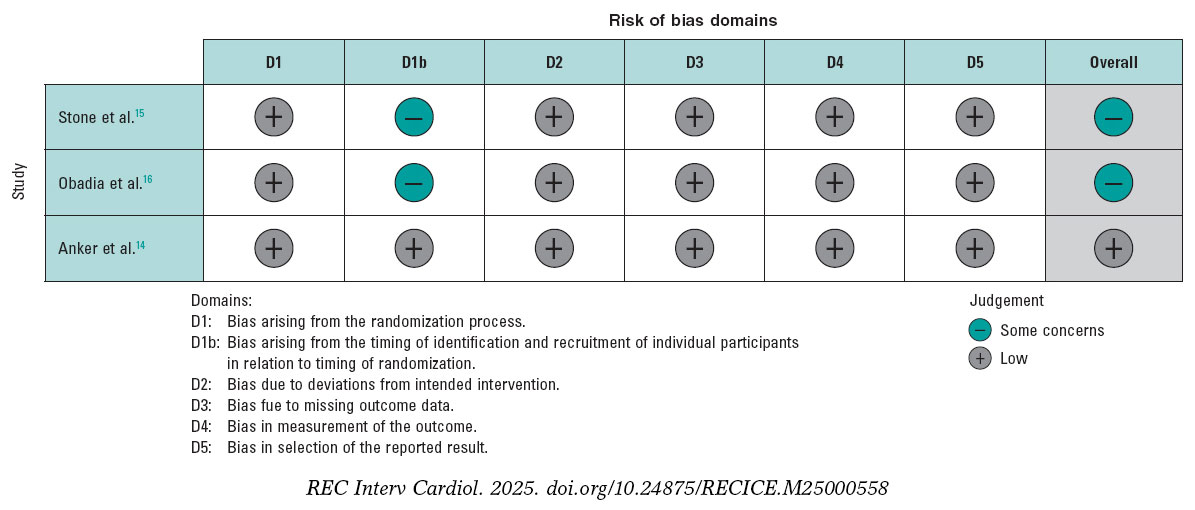
Figure 2. Risk of bias for each included randomized clinical trial. The bibliographical references mentioned in this figure correspond to Stone et al.,14 Obadia et al.,15 and Anker et al.16.
Outcomes
HF-related hospitalization
The analysis of this outcome showed a significant reduction, with a RR of 0.71 (95%CI, 0.56-0.90; P = .004). However, substantial heterogeneity was observed, with an I² value of 79%. Considering the differences in the MITRA-FR study population, we conducted an exploratory analysis excluding this trial. Results still demonstrated a significant reduction in the risk of hospitalization, with a RR of 0.63 (95%CI, 0.55-0.72, P < .00001), along with a marked improvement in heterogeneity (I² = 0%). Of note, this finding is exploratory and should be interpreted with caution.
The analysis, including all studies, is shown in figure 3A, and the exploratory analyses are shown in figure 3B. Furthermore, we analyzed HR for this endpoint and, given the limited number of studies, we conducted a sensitivity analysis using the Hartung-Knapp-Sidik-Jonkman (HKSJ) method; all these analyses are provided in the supplementary data (figures S1-S4).
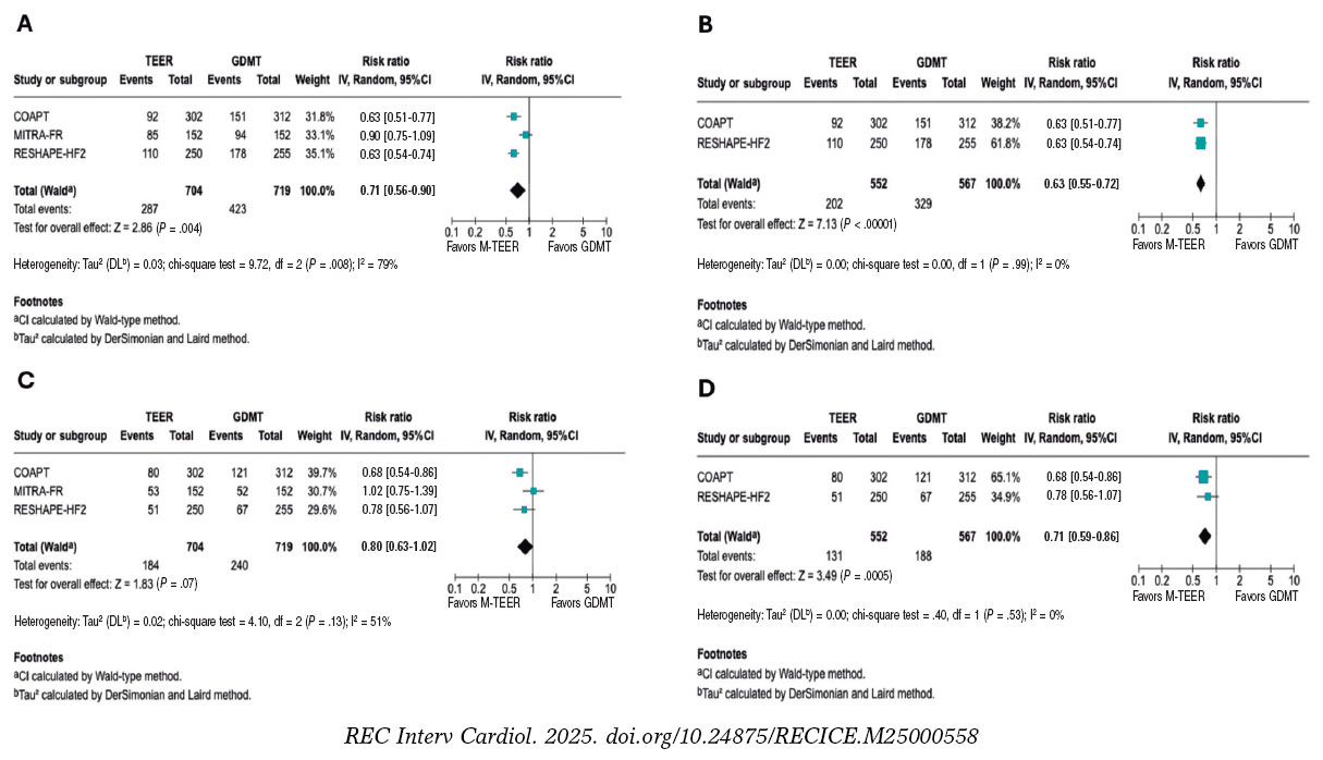
Figure 3. Forest plot of risk ratios (RR). Lines denote the 95% confidence intervals (95%CI) for each trial. A: forest plot of RR for HF related hospitalization. B: forest plot of exploratory RR for HF-related hospitalization. C: forest plot of RR for all-cause mortality. D: forest plot of exploratory RR for all-cause mortality. 95%CI, 95% confidence interval; GDMT, guideline directed medical therapy; M-TEER, mitral transcatheter edge-to-edge repair. The bibliographical references mentioned in this figure correspond to Stone et al.,14 Obadia et al.,15 and Anker et al.16.
All-cause mortality
The RR for all-cause mortality was 0.80 (95%CI, 0.63–1.02; P = .07). However, substantial heterogeneity was observed across the studies (I² = 56%). In the exploratory analysis, a significant reduction in mortality was found, with an RR of 0.71 (95%CI, 0.59–0.86; P = .0005) and no heterogeneity across the studies (I² = 0%). The analyses, including all studies, are shown in figure 3C, and the exploratory analysis in figure 3D.
Both the HR analysis and the sensitivity analysis are provided in the supplementary data (figures S5-S8).
Cardiac death
The RR for death from cardiovascular causes was 0.81 (95%CI, 0.62–1.06, P = .12), with low to moderate heterogeneity (I² = 48%). In the exploratory analysis, a significant reduction in cardiac death was observed, with an RR of 0.74 (95%CI, 0.55–1.00; P = .05) and low heterogeneity (I² = 41%). The analyses, including all studies, are shown in figure 4A, while the exploratory analysis, in figure 4B. The sensitivity analysis and HR results are provided in the supplementary data (figures S9-S12).
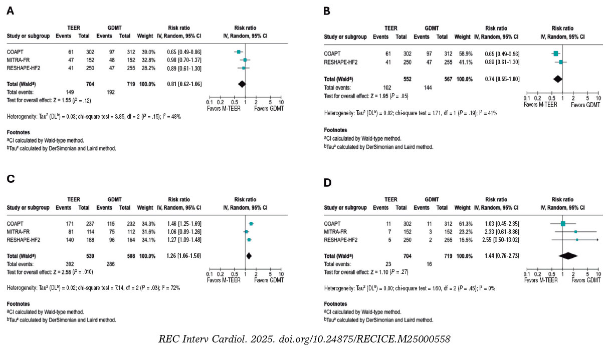
Figure 4. Forest plot of risk ratio (RR). A: forest plot of RR for cardiac death. B: forest plot of exploratory RR for cardiac death. C: forest plot of RR of NYHA FC I/II at 1 year. D: forest plot of RR for stroke. 95%CI, 95% confidence interval; GDMT, guideline directed medical therapy; M-TEER, mitral transcatheter edge-to-edge repair. The bibliographical references mentioned in this figure correspond to Stone et al.,14 Obadia et al.,15 and Anker et al16. Lines denote the 95%CI for each trial.
NYHA FC
In this analysis of the NYHA FC, we observed that patients undergoing M-TEER are more likely to be found in NYHA FC I/II at 12 months, with a RR 1.26 (95%CI, 1.06–1.50; P = .010), respectively. Of note, the high heterogeneity (I² = 72%). Furthermore, it is also essential to note that the NYHA FC is a subjective classification, which is why the findings of this outcome should be interpreted with caution. These results are shown in figure 4C.
Stroke
There were no statistical differences between M-TEER and optimal medical therapy regarding stroke, with a RR of 1.44 (95%CI, 0.76 - 2.73, P = .27) without any heterogeneity being reported across the studies (I² = 0%). These results are shown in figure 4D.
Myocardial Infarction
Regarding the risk of myocardial infarction, our analysis demonstrated no statistical difference between the M-TEER and optimal medical therapy groups (RR, 0.83; 95%CI, 0.43-1.61; P = .58). No heterogeneity among the studies was observed (I² = 0%); this finding is shown in supplementary data (figure S13).
Exploratory analysis by severity
We conducted an exploratory analysis stratified by the baseline grade of mitral regurgitation. Data on the composite endpoint of all-cause death or HF-related hospitalization were available according to MR grade. For patients with MR 3+, the RR was 0.75 (95%CI, 0.56-1.00; P = .05; I² = 12%). For those with severe MR 4+, data were available only from the RESHAPE-HF2 and COAPT trials, yielding a RR of 0.55 (95%CI, 0.42-0.73; P = .0001; I² = 0%). This finding is shown in the supplementary data (figure S14 to figure S15).
DISCUSSION
Our meta-analysis provides an evaluation of 3 major RCTs, COAPT15, MITRA-FR16, and RESHAPE-HF214, evaluating M-TEER plus GDMT vs GDMT alone in patients with secondary MR. In patients with persistent symptoms despite adequate GDMT and who do not meet the criteria for definitive surgical replacement, mitral M-TEER has emerged as a promising alternative. In this updated meta-analysis, no statistically significant effect on all-cause mortality was found; however, the marked between-trial heterogeneity suggests that M-TEER provides meaningful clinical benefit when used in appropriately selected patients.
Our study showed that heterogeneity of results was mainly driven by MITRA-FR. COAPT and RESHAPE-HF2 have multiple similar differences compared with MITRA-FR, as evidenced in trial cohort baseline characteristics, methods, and outcome directions. This is better portrayed by an exploratory analysis comparing COAPT and RESHAPE-HF2 results, showing a 37% reduction in the risk of HF-related hospitalization risk and a 32% reduction in cardiac death (figure 3). The overall neutral mortality rate resulting from our study may be driven by heterogeneity across trials rather than a lack of intrinsic therapeutic benefit.
Among the main differences across trials, the MITRA-FR trial had broader inclusion of patients with tricuspid regurgitation, advanced LV remodeling, greater dilation, and smaller EROA vs the other 2 trials. Another key source of heterogeneity was baseline MR severity. COAPT and RESHAPE-HF2 primarily enrolled patients with grade 3+ and 4+ MR, whereas MITRA-FR is only focused on grade 4+, which may contribute to the observed outcome differences. In our exploratory analysis by MR grade, data showed consistent trends favoring M-TEER; however, these findings should be interpreted with caution due to limited subgroup data.
As proposed by Paul et al.,19 one strategy to evaluate secondary MR is to consider the proportion between EROA and LVEDV. In the MITRA-FR trial, many patients had smaller EROA with markedly dilated ventricles, a phenotype in which GDMT may have more impact than valve proceduren. In the COAPT and RESHAPE-HF2 trials, a greater proportion of patients had large EROA relative to the LVEDV,20 making MR a primary driver of symptoms and outcomes, and, therefore, with potential for greater benefit from M-TEER.
In the trial-level subgroup analyses, the COAPT15 and RESHAPE-HF214 trials reported no significant interaction between treatment assignment and ischemic vs non-ischemic etiology, which indicates consistency of benefit across etiologies. Similarly, the MITRA-FR16 did not identify significant heterogeneity regarding etiology across prespecified subgroups. This suggests that ischemic vs non-ischemic alone should not be used to decide the treatment option in secondary MR. Instead, clinical decisions should prioritize other factors, such as the MR severity, the degree of LV dysfunction, symptom burden, and response to optimal medical therapy.21
Furthermore, differences between trials in procedural success are noteworthy, defined as achieving MR ≤ 2+. This was substantially lower in the MITRA-FR (75.6% MR ≤ 2+ at discharge) vs the COAPT (94.8% MR ≤ 2+ at 12 months) and the RESHAPE-HF2 (90.4% MR ≤ 2+ at 12 months). Multiple reasons could have influenced such differences; for instance, operator experience requirement in MITRA-FR (≥ 5 prior MitraClip cases) vs the high-volume and more experienced centers from the other trials. Similarly, post-approval data from the U.S. SSTS/ACC TVT Registry22 demonstrated > 90% MR reduction to ≤ 2+ and survival rates consistent with trial outcomes, underscoring the reproducibility of clinical benefit in experienced centers. Furthermore, device technology is a main contributor to these differences, from first-generation clips in MITRA-FR, to second-generation in COAPT, to the fourth-generation in RESHAPE-HF2 with independent leaflet grasping and wider arm options, potentially explaining discrepant clinical results regarding MR durability and outcomes. Lastly, GDMT implementation differed across trials. MITRA-FR was conducted before the widespread use of ARNI and SGLT2 inhibitors; COAPT was conducted before the adoption of SGLT2 inhibitors; and RESHAPE-HF2 reflects the contemporary use of quadruple therapy.
Despite the differences in patient phenotypes across the trials, in many developing countries, access to transcatheter valve procedures remains limited, primarily due to their high cost and the need for specialized infrastructure.23,24 The pronounced disparities in access to cutting-edge technologies, coupled with the centralization of these procedures in a few urban centers or high-specialty hospitals, a phenomenon observed even in high-income countries such as the United States, leave a substantial portion of the population without viable treatment options.25 Consequently, the most impactful and broadly deployable intervention in these regions remains rigorous GDMT optimization and provider education. Nevertheless, patient phenotyping should still be performed to identify those who may potentially have an outsized benefit from future M-TEER referrals.
By integrating trial-level population characteristics with clinical outcomes, our work bridges the gap between isolated trial findings and real-world patient selection, thus offering a potential framework for future prospective studies and for refining guideline criteria. While the results from our study must be interpreted with caution, they provide hypothesis-generating evidence supporting the concept that patient selection, particularly considering the balance between EROA and LVEDV and GDMT optimization, may be critical to optimizing the benefit of M-TEER in secondary MR. This approach moves beyond the broad application of current guideline recommendations and points toward a more individualized strategy in which anatomical and functional parameters guide intervention.
Limitations
The primary limitation of this study lies in the heterogeneity of the populations enrolled in the randomized controlled trials, as evidenced by the I2 observed in the analyses, which may influence the overall results. Second, differences in MR severity across trials represent a key limitation. While the COAPT and RESHAPE-HF2 trials mainly included patients with MR grade 3+ or 4, MITRA-FR enrolled a more severe grade, which may partly explain divergent results. Lastly, an individual patient-level meta-analysis could not be conducted due to lack of data; this level of analysis would further increase statistical power, especially in subgroup analyses, enhancing the robustness and generalizability of the findings.
CONCLUSIONS
In this meta-analysis, M-TEER plus GDMT shows a lower risk of HF-related hospitalization vs GDMT alone. We did not find any differences in the risk of all-cause mortality, cardiac death, stroke, or myocardial infarction.
FUNDING
None declared.
ETHICAL CONSIDERATIONS
This meta-analysis was performed using data from previously published studies. As it is based entirely on secondary data, no new data were collected from human or animal participants; on the other hand, the use of SAGER guidelines was not applicable in this study. All included studies were approved from the relevant center ethics committees. The authors confirm that all data utilized were publicly accessible, and no confidential information was used without proper authorization.
STATEMENT ON THE USE OF ARTIFICIAL INTELLIGENCE
During the preparation of this work, the authors used ChatGPT 4o to review the document’s syntax and grammar. After using this tool/service, the authors reviewed and edited the content as needed and take full responsibility for the content of the published article.
AUTHORS’ CONTRIBUTIONS
D. Paulino-González: conceptualization; formal analysis, writing, review, and editing. A.L. García-Loera: methodology, investigation, writing, review, and editing. D.A. Navarro-Martínez: methodology, formal analysis. M.A. Pardiño-Vega: writing, review, and editing, supervision. K.P. Zúñiga-Montaño: investigation, writing, review, and editing.
CONFLICTS OF INTEREST
None declared.
WHAT IS KNOWN ABOUT THE TOPIC?
- Mitral regurgitation is a prevalent condition that negatively affects the patients’ quality of life. M-TEER-has emerged as an alternative therapeutic option for patients who are ineligible for surgery. However, its benefit remains unclear vs GDMT, as clinical trials evaluating this procedure have reported disparate results, with benefit in primary outcomes observed in the COAPT trial and in the recently published RHESAPE-HF2 trial, but not in the MITRA-FR trial; discrepancy in the results requires further study.
WHAT DOES THIS STUDY ADD?
- This meta-analysis provides a comprehensive evaluation of the evidence comparing M-TEER plus GDMT vs GDMT alone in secondary mitral regurgitation. Our findings confirm that M-TEER is associated with a significant reduction in HF-related hospitalizations. Of note, by examining trial populations in detail, we highlight how patient selection influences outcomes: in studies enrolling patients with less ventricular dilation and lower biomarker levels of congestion (such as the COAPT and RESHAPE-HF2 trials), M-TEER was associated with additional benefits in cardiac death and all-cause mortality. These results underscore the potential role of anatomical and functional parameters, such as the balance between EROA and LVEDV, in identifying patients most likely to benefit from this procedure.
SUPPLEMENTARY DATA
REFERENCES
1. Chehab O, Roberts-Thomson R, Ng Yin Ling C, et al. Secondary mitral regurgitation:pathophysiology, proportionality and prognosis. Heart. 2020;106:716-723.
2. Sengodan P, Younes A, Shah N, Maraey A, Chitwood WR Jr, Movahed A. Contemporary review of the evolution of various treatment modalities for mitral regurgitation. Expert Rev Cardiovasc Ther. 2024;22:639-651.
3. Nishimura RA, Otto CM, Bonow RO, et al. 2017 AHA/ACC Focused Update of the 2014 AHA/ACC Guideline for the Management of Patients With Valvular Heart Disease:A Report of the American College of Cardiology/American Heart Association Task Force on Clinical Practice Guidelines. Circulation. 2017;135:e1159-e1195.
4. Itabashi Y, Kobayashi S, Mizutani Y, Torikai K, Taguchi I. Treatment of secondary mitral regurgitation by transcatheter edge-to-edge repair using MitraClip. J Med Ultrason (2001). 2022;49:389-403.
5. Barnes C, Sharma H, Gamble J, Dawkins S. Management of secondary mitral regurgitation:from drugs to devices. Heart. 2024;110:1099-1106.
6. Page MJ, McKenzie JE, Bossuyt PM, et al. The PRISMA 2020 statement:an updated guideline for reporting systematic reviews. BMJ. 2021;372:n71.
7. Sterne JAC, Savovic´J, Page MJ, et al. RoB 2:a revised tool for assessing risk of bias in randomised trials. BMJ. 2019;366:l4898.
8. Heidenreich PA, Bozkurt B, Aguilar D, et al. 2022 AHA/ACC/HFSA Guideline for the Management of Heart Failure:A Report of the American College of Cardiology/American Heart Association Joint Committee on Clinical Practice Guidelines [published correction appears in Circulation. 2022;145:e1033.] [published correction appears in Circulation. 2022;146:e185.] [published correction appears in Circulation. 2023;147:e674]. Circulation. 2022;145:e895-e1032.
9. Abraham WT, Psotka MA, Fiuzat M, et al. Standardized Definitions for Evaluation of Heart Failure Therapies:Scientific Expert Panel From the Heart Failure Collaboratory and Academic Research Consortium. JACC Heart Fail. 2020;8:961-972.
10. Sacco RL, Kasner SE, Broderick JP, et al. An updated definition of stroke for the 21st century:a statement for healthcare professionals from the American Heart Association/American Stroke Association [published correction appears in Stroke. 2019;50:e239]. Stroke.2013;44:2064-2089.
11. Thygesen K, Alpert JS, Jaffe AS, et al. Fourth Universal Definition of Myocardial Infarction (2018). Glob Heart. 2018;13:305-338.
12. Higgins JP, Thompson SG, Deeks JJ, Altman DG. Measuring inconsistency in meta-analyses. BMJ. 2003;327:557-560.
13. DerSimonian R, Laird N. Meta-analysis in clinical trials. Control Clin Trials. 1986;7:177-188.
14. Anker SD, Friede T, von Bardeleben RS, et al. Transcatheter Valve Repair in Heart Failure with Moderate to Severe Mitral Regurgitation. N Engl J Med.2024;391:1799-1809.
15. Stone GW, Lindenfeld J, Abraham WT, et al. Transcatheter Mitral-Valve Repair in Patients with Heart Failure. N Engl J Med. 2018;379:2307-2318.
16. Obadia JF, Messika-Zeitoun D, Leurent G, et al. Percutaneous Repair or Medical Treatment for Secondary Mitral Regurgitation. N Engl J Med.2018;379:2297-2306.
17. Iung B, Armoiry X, Vahanian A, et al. Percutaneous repair or medical treatment for secondary mitral regurgitation:outcomes at 2 years. Eur J Heart Fail. 2019;21:1619-1627.
18. Ponikowski P, Friede T, von Bardeleben RS, et al. Hospitalization of Symptomatic Patients With Heart Failure and Moderate to Severe Functional Mitral Regurgitation Treated With MitraClip:Insights From RESHAPE-HF2. J Am Coll Cardiol. 2024;84:2347-2363.
19. Grayburn PA, Sannino A, Packer M. Proportionate and Disproportionate Functional Mitral Regurgitation:A New Conceptual Framework That Reconciles the Results of the MITRA-FR and COAPT Trials. JACC Cardiovasc Imaging. 2019;12:353-362.
20. Anker SD, Friede T, von Bardeleben RS. Percutaneous repair of moderate-to-severe or severe functional mitral regurgitation in patients with symptomatic heart failure:Baseline characteristics of patients in the RESHAPE-HF2 trial and comparison to COAPT and MITRA-FR trials. Eur J Heart Fail. 2024;26:1608-1615.
21. Nappi F, Singh SSA, Bellomo F.Exploring the Operative Strategy for Secondary Mitral Regurgitation:A Systematic Review. Biomed Res Int.2021;2021:3466813.
22. American College of Cardiology. STS/ACC TVT Registry Analysis Finds TMVr Safe and Effective in Real-World Setting. ACC. 2023 Mar 5. Available at: https://www.acc.org/Latest-in-Cardiology/Articles/2023/03/01/22/45/Sun-1215pm-sts-acc-tvt-acc-2023. Accessed 1 Jul 2025.
23. Bernardi FLM, Ribeiro HB, Nombela-Franco L, et al. Recent Developments and Current Status of Transcatheter Aortic Valve Replacement Practice in Latin America - the WRITTEN LATAM Study. Arq Bras Cardiol. 2022;118:1085-1096.
24. Bana A. TAVR-present, future, and challenges in developing countries. Indian J Thorac Cardiovasc Surg. 2019;35:473-484.
25. Steitieh D, Zaidi A, Xu S, et al. Racial Disparities in Access to High-Volume Mitral Valve Transcatheter Edge-to-Edge Repair Centers. J Soc Cardiovasc Angiogr Interv. 2022;1:100398.


Review Articles

Interviews

An interview with Bruno Scheller
aServicio de Cardiología, Hospital Universitario de La Princesa, Instituto de Investigación Sanitaria de La Princesa (IIS-IP), Universidad Autónoma de Madrid, Spain
bCentro de Investigación Biomédica en Red de Enfermedades Cardiovasculares (CIBERCV), Instituto de Salud Carlos III, Madrid, Spain


