Article
Adult congenital heart disease
REC Interv Cardiol. 2022;4:173-180
Spanish cardiac catheterization in congenital heart diseases registry. First official report from the ACI-SEC and the GTH-SECPCC (2020)
Registro Español de Intervencionismo en Cardiopatías Congénitas. Primer Informe Oficial de la ACI-SEC y el GTH-SECPCC (2020)
aServicio de Cardiología Pediátrica, Hospital General Universitario Gregorio Marañón, Madrid, Spain
bServicio de Cardiología Pediátrica, Hospital Universitario Virgen del Rocío, Sevilla, Spain
cServicio de Cardiología, Hospital Universitari de Bellvitge, IDIBELL, Universitat de Barcelona, L’Hospitalet de Llobregat, Barcelona, Spain
dServicio de Cardiología Infantil, Hospital Universitario La Paz, Madrid, Spain
eDepartamento de Cardiología, Hospital Clínico Universitario de Valladolid, Valladolid, Spain
fCentro de Investigación Biomédica en Red de Enfermedades Cardiovasculares (CIBERCV), Spain
gServicio de Cardiología Pediátrica, Hemodinámica Pediátrica, Hospital Universitari Vall d’Hebron, Barcelona, Spain
hSección de Hemodinámica-Cardiología, Hospital Universitario de Cruces, Barakaldo, Bilbao, Spain
iServicio de Cardiología Pediátrica y Unidad de Cardiopatías Congénitas, Hospital Universitario Ramón y Cajal, Madrid, Spain
jInstituto Pediátrico del Corazón, Hospital Universitario 12 de Octubre, Instituto de Investigación Sanitaria 12 de Octubre (imas12), Madrid, Spain
kSección de Hemodinámica-Cardiología, Hospital Universitario Son Espases, Palma de Mallorca, Spain
lSección de Cardiología Infantil, Hospital Universitario y Politécnico La Fe, Valencia, Spain
mSección de Cardiología Infantil, Hospital Miguel Servet, Zaragoza, Spain
nSección de Cardiología Pediátrica, Hospital Universitario Virgen de la Arrixaca, El Palmar, Murcia, Spain
oServicio de Cardiología, Hospital Universitario Reina Sofía, Instituto Maimónides de Investigación Biomédica (IMIBIC), Universidad de Córdoba, Córdoba, Spain
pUnidad de Cardiopatías Congénitas, Servicio de Pediatría, Complejo Hospitalario Universitario de A Coruña, A Coruña, Spain
qServicio de Cardiología, Hospital Universitario Virgen de las Nieves de Granada, Granada, Spain
rServicio de Cardiología, Hospital General Universitario Gregorio Marañón, Instituto de Investigación Sanitaria Gregorio Marañón (IiSGM), Universidad Complutense de Madrid, Madrid, Spain
sSección de Cardiología Pediátrica, Hospital Regional Universitario de Málaga, Málaga, Spain
RESUMEN
Introduction and objectives: The Interventional Cardiology Association of the Spanish Society of Cardiology (ACI-SEC) and the Interventional Working Group of the Spanish Society of Pediatric Cardiology (GTH-SECPCC) present their 2023 annual activity report.
Methods: Participation was open to all Spanish centers with cath labs and interventional programs for congenital heart disease. Data was collected online and analyzed by an external company, and members from the ACI-SEC and the GTH-SECPCC.
Results: A total of 19 centers participated (15 public and 4 private). Interventional data on adult congenital diseases was contributed to the Registry of Hemodynamics and Interventional Cardiology of the ACI-SEC in 2023 by another 114 hospitals and, then, added to the analysis. A total of 1127 diagnostic studies (1.2% less than 2022) and 3856 interventional cardiac catheterizations (53.7% more than 2022) were registered, 2439 of which (65%) were performed on patients older than 18 years. The most widely performed procedures were foramen ovale closure (1498 cases), atrial septal defect closure (700 cases), transcatheter closure of the patent ductus arteriosus (344 cases) and aortic coarctation (221 cases). The most significant volume increment of cases was associated with ventricular septal defect closure (181%), foramen ovale closure (87%), and aortic coarctation (75%). Interventional procedures were considered successful in 98.3% of the cases, with rates of major procedural complication and in-hospital mortality of 1% and 0.05%, respectively.
Conclusions: We present the fourth Spanish cardiac catheterization in congenital heart diseases registry with data from 2023. We observed a significant rise in interventional procedures, notably ventricular septal defect and patent foramen ovale closures. Most interventional techniques continue to demonstrate excellent safety and efficacy outcomes.
Keywords: Congenital heart disease. Cardiac catheterization. Ventricular septal defect closure. Transcatheter aortic valve implantation.
ABSTRACT
Introducción y objetivos: La Asociación de Cardiología Intervencionista de la Sociedad Española de Cardiología (ACI-SEC) y el Grupo de Trabajo de Hemodinámica de la Sociedad Española de Cardiología Pediátrica y Cardiopatías Congénitas (GTH-SECPCC) presentan su informe anual de actividad hemodinámica en cardiopatías congénitas correspondiente al año 2023.
Métodos: Se invitó a participar a los centros españoles con laboratorio de hemodinámica y actividad intervencionista en cardiopatías congénitas. La recogida de datos fue telemática y su análisis lo realizó una empresa externa junto con miembros de la ACI-SEC y el GTH-SECPCC.
Resultados: Participaron en el registro 19 centros (15 públicos y 4 privados). Se incorporaron al análisis los datos de intervencionismo en cardiopatías congénitas del adulto aportados por otros 114 hospitales al Registro de Hemodinámica y Cardiología Intervencionista de la ACI-SEC del año 2023. Se registraron 1.127 estudios diagnósticos (1,2% menos que en 2022) y 3.856 cateterismos intervencionistas (53,7% más que en 2022), de los que 2.439 (65%) se realizaron en mayores de 18 años. Las técnicas más frecuentes fueron los cierres de foramen oval (1.498 casos), de comunicación interauricular (700 casos) y de ductus arterioso (344 casos), y la coartación de aorta (221 casos). Respecto al año anterior, se incrementaron significativamente las técnicas de cierre de comunicación interventricular (181%), cierre de foramen oval (87%) y coartación de aorta (75%). La tasa de éxito en los procedimientos intervencionistas fue del 98,3%, con una tasa de complicaciones mayores del 1% y una mortalidad intrahospitalaria del 0,05%.
Conclusiones: Se presenta el informe del Registro español de intervencionismo en cardiopatías congénitas del año 2023. Se ha comunicado un aumento muy significativo de los procedimientos terapéuticos, destacando el incremento en los cierres de comunicación interventricular y de foramen oval. Todas las técnicas intervencionistas han mostrado excelentes datos de seguridad y eficacia.
Palabras clave: Cardiopatías congénitas. Cateterismo cardiaco. Cierre de comunicación interventricular. Implante percutáneo de válvula aórtica.
INTRODUCTION
Over the past 5 years, the collaboration between the Interventional Cardiology Association of the Spanish Society of Cardiology (ACI-SEC) and the Interventional Working Group of the Spanish Society of Pediatric Cardiology (GTH-SECPCC) has led to the consolidation of the Spanish cardiac catheterization in congenital heart diseases registry, substantiated thus far by the publication of its first 3 reports on the activities conducted in 2020,1 2021,2 and 20223 (figure 1). The first 2 reports demonstrated that the number of centers participating in the registry, although highly representative of pediatric activity, did not accurately reflect the activity of adult congenital heart disease conducted in Spain.4,5,6 Therefore, the analysis of the current report presented in this article, on the activity conducted in 2023, has incorporated—same as in the previous report—the interventional activity in congenital heart disease from the Registry of Hemodynamics and Interventional Cardiology of the ACI-SEC for that same year.7 This methodology has proven to more reliably quantify hemodynamic procedures in congenital heart disease across all age groups. The findings from this edition were presented on June 14, 2024, at the ACI-SEC Congress held in Las Palmas de Gran Canaria (Canary Islands, Spain).
Figure 1. Comparison of the number of interventional procedures in 2020, 2021, 2022, and 2023.
METHODS
Data come from an annually updated, retrospective, voluntary, and non-audited registry. The inclusion of interventional data on congenital heart disease from the Registry of Hemodynamics and Interventional Cardiology of the ACI-SEC was possible due to the harmonization of questionnaires between the 2 registries conducted the previous year, which continued to undergo improvements.
All hospitals already participating in the Registry of Hemodynamics and Interventional Cardiology of the ACI-SEC, and all hospitals represented in the GTH-SECPCC, were asked to participate. Data was collected through an electronic database managed by an external company (pInvestiga España), which analyzed the registry results and compared them with those from previous years in collaboration with members of the GTH-SECPCC and the ACI-SEC board. Discrepant or particularly noteworthy data were cleaned and verified with the responsible investigator.
Due to the methodological characteristics of the study and the fact that it is only an activity registry, both the approval from the ethics committee and the processing of informed consent were deemed unnecessary.
RESULTS
Resources and infrastructure
A total of 19 hospitals participated (3 less than in 2021), of which 15 belong to the public health care system and 4 to the private sector (table 1 of the supplementary data). In addition, the analysis incorporated data on adult congenital heart disease interventions from another 114 hospitals (15 more than in 2022) included in the 2023 Registry of Hemodynamics and Interventional Cardiology of the ACI-SEC (table 2 of the supplementary data).
Table 1. Number of interventional procedures and distribution by age groups
| Variable | Fetal | < 1 month | 1 month to 1 year | 1-18 years | > 18 years | Total |
|---|---|---|---|---|---|---|
| Interventional procedures | 4 (0.1) | 165 (4.2) | 281 (7.2) | 967 (25) | 2439 (63.2) | 3856 |
| Congenital aortic valvuloplasty | 2 (2.5) | 18 (22.5) | 16 (20.0) | 24 (30.0) | 20 (25.0) | 80 |
| Congenital pulmonary valvuloplasty | 2 (1.2) | 43 (25.9) | 44 (26.5) | 19 (11.4) | 58 (34.9) | 166 |
| Congenital mitral valvuloplasty | – | 0 | 0 | 1 (5.2) | 18 (94.7) | 19 |
| Pulmonary angioplasty | – | 13 (12.0) | 8 (7.4) | 63 (58.3) | 24 (22.2) | 108 |
| Pulmonary branch angioplasty | – | 5 (2.2) | 48 (21.8) | 109 (49.5) | 58 (26.3) | 220 |
| Aortic coarctation | – | 7 (3.1) | 34 (15.3) | 69 (31.2) | 111 (50.2) | 221 |
| Other angioplasties | – | 28 (21.3) | 17 (12.9) | 63 (48.0) | 23 (17.5) | 131 |
| Patent foramen ovale closure | – | 0 | 0 | 12 (0.8) | 1486 (99.1) | 1498 |
| Atrial septal defect closure | – | 0 | 7 (1.0)a | 238 (34.0) | 455 (65.0) | 700 |
| Ductus arteriosus closure | 15 (4.3)b | 20 (5.8)b | 38 (11.1)b | 208 (60.4) | 63 (18.3) | 344 |
| Ventricular septal defect closure | – | 0 | 9 (9.6) | 58 (62.3) | 40 (37.9) | 107 |
| Other occlusions | – | 2 (2.3) | 17 (20.0) | 45 (52.9) | 21 (24.7) | 85 |
| Foreign body removal | – | 1 (5.0) | 3 (15.0) | 15 (75.0) | 1 (5.0) | 20 |
| Atrial septostomy and transseptal puncture | 0 | 48 (75.0) | 5 (7.8) | 9 (14.0) | 2 (3.1) | 64 |
| Transcatheter aortic valve implantation | – | 0 | 0 | 34 (36.5)c | 59 (63.4) | 93 |
|
Data are expressed as no. (%). a In this case, < 1 month and 1 month to 1 year are not collected separately, so the value corresponds to < 1 year. bFor ductus arteriosus closure, the groups are premature (fetal), < 6 months (< 1 month) and 6 months to 1 year (1 month to 1 year). cCollected as < 18 years, so the value corresponds to < 18 years. |
||||||
Table 2. Number of interventional catheterizations in patients > 18 years and distribution according to origin registry
| Interventional procedures | > 18 years | ||
|---|---|---|---|
| Total | RICCa | RHCIb | |
| Congenital aortic valvuloplasty | 20 | 1 (5.0) | 19 (95.0) |
| Congenital pulmonary valvuloplasty | 58 | 22 (37.9) | 36 (62.0) |
| Congenital mitral valvuloplasty | 18 | 1 (5.5) | 17 (94.5) |
| Pulmonary angioplasty | 24 | 24 (100.0) | 0 (0.0) |
| Pulmonary branch angioplasty | 58 | 30 (51.7) | 28 (48.3) |
| Aortic coarctation | 111 | 29 (26.1) | 82 (73.8) |
| Other angioplasties | 23 | 23 (100.0) | 0 (0.0) |
| Patent foramen ovale closure | 1486 | 149 (10.0) | 1337 (90.0) |
| Atrial septal defect closure | 455 | 53 (11.6) | 402 (88.4) |
| Ductus arteriosus closure | 63 | 3 (4.7) | 60 (95.2) |
| Ventricular septal defect closure | 40 | 5 (12.5) | 35 (87.5) |
| Other occlusions | 21 | 21 (100.0) | 0 (0.0) |
| Foreign body removal | 1 | 1 (100.0) | 0 (0.0) |
| Atrial septostomy and transseptal puncture | 2 | 2 (100.0) | 0 (0.0) |
| Transcatheter aortic valve implantation | 59 | 59 (100.0) | 0 (0.0) |
| Total | 2439 | 423 (17.3) | 2016 (82.7) |
|
RHCI, Registry of Hemodynamics and Interventional Cardiology of the ACI-SEC; RICC, Spanish cardiac catheterization in congenital heart diseases registry of the ACI-SEC. Data are expressed as no (%). aData provided by the 19 centers participating to the 2023 Interventional Cardiology in Congenital Heart Diseases Registry of the ACI-SEC and the GTH-SECPCC. bData provided by 96 centers to the 2023 Registry of Hemodynamics and Interventional Cardiology of the ACI-SEC. |
|||
A total of 38 cath labs performing interventional procedures in congenital heart disease were registered, 7 of which (18.4%) only deal with pediatric procedures; 9 are equipped with biplane systems and 8 implement rotational angiography. The median number of monthly days dedicated to congenital heart disease interventions per hospital was 7 (range, 3–18) vs 6 days in 2022. Thirteen centers (68.4%) provide 24-hour emergency interventional care, including services for pediatric patients.
Regarding medical staff, a total of 68 interventional cardiologists were dedicated to this activity, including 36 (52.9%) specialized in adult interventions and 32 (47.1%) in pediatric interventions.
Diagnostic procedures
A total of 1127 diagnostic studies were recorded, representing a 1.2% decrease compared with the previous year. The age distribution was as follows: 27 (2.4%) were performed in infants younger than 1 month, 95 (8.4%) in children aged 1 month to 1 year, 548 (48.6%) in patients aged 1–18 years, and 457 (40.6%) in patients older than 18 years.
A total of 70 procedures (6.5%) were categorized as emergencies. Regarding morbidity, there were 5 cases (0.4%) of severe complications: 2 vascular events, 1 arrhythmia with severe hemodynamic instability and cardiorespiratory arrest, 1 anaphylactic reaction, and 1 neurological event. There were no procedural deaths.
Interventional procedures
Reported activity increased by 53.7% compared with the previous year for a total of 3856 therapeutic catheterizations registered and categorized into 15 different groups. Their case distribution and age breakdown are shown in table 1. Of the 2439 procedures (63.2%) performed in patients older than 18 years, 2016 (82.7%) came from data added from the Registry of Hemodynamics and Interventional Cardiology of the ACI-SEC (table 2).
A total of 195 cardiac catheterizations were categorized as emergencies (10.9% of all procedures for which this information was available). The volume of interventional procedures per center was as follows: 7 hospitals (36.8%) recorded > 150 cardiac catheterizations; 3 hospitals (15.7%) between 75 and 150 procedures, and 9 (47.3%) < 75 procedures. The overall effectiveness reported for the different interventional techniques was 97.9%, with most procedures showing success rates exceeding 95% (table 3).
Table 3. Summary of reported efficacy of interventional procedures
| Interventional procedures | n | n with success or ineffectiveness data |
Success | Ineffective |
|---|---|---|---|---|
| Congenital aortic valvuloplasty | 80 | 65 (81.2) | 59 (90.7) | 6 (9.2) |
| Congenital pulmonary valvuloplasty | 166 | 133 (80.1) | 129 (96.9) | 2 (1.5) |
| Congenital mitral valvuloplasty | 19 | 19 (100) | 18 (94.7) | 1 (5.3) |
| Pulmonary angioplasty | 108 | 89 (82.4) | 81 (91.0) | 8 (8.9) |
| Pulmonary branch angioplasty | 220 | 193 (87.7) | 181 (93.7) | 12 (6.2) |
| Aortic coarctation | 221 | 139 (62.8) | 131 (94.2) | 8 (5.7) |
| Other angioplasties | 131 | 131 (100) | 128 (96.9) | 4 (3.0) |
| Patent foramen ovale closure | 700 | 547 (78.4) | 539 (98.5) | 8 (1.4) |
| Atrial septal defect closure | 1498 | 1188 (79.3) | 1182 (99.4) | 6 (0.5) |
| Ductus arteriosus closure | 344 | 251 (72.9) | 249 (99.2) | 3 (1.1) |
| Ventricular septal defect closure | 107 | 81 (75.7) | 78 (96.2) | 3 (3.7) |
| Other occlusions | 85 | 85 (100) | 85 (100.0) | 0 (0.0) |
| Foreign body removal | 20 | 20 (100) | 20 (100.0) | 0 (0.0) |
| Atrial septostomy and transseptal puncture | 64 | 64 (100) | 63 (98.4) | 1 (1.5) |
| Transcatheter aortic valve implantation | 93 | 92 (98.9) | 91 (98.9) | 1 (1.1) |
| Total | 3856 | 3084 (79.9) | 3034 (98.3) | 63 (2.0) |
|
Data are expressed as no (%). Percentage of success or ineffectiveness based on the number of procedures with available data. |
||||
Transcatheter valvuloplasties
A total of 80 aortic valvuloplasties were reported for congenital aortic stenosis, reflecting a 19.4% increase from 2022. Two of these procedures were performed in fetuses. Sixty cases (75%) involved patients younger than 18 years, and in 77% of procedures, the dilation was performed on native, previously untreated valves.
A total of 166 pulmonary valvuloplasties were performed, marking a 20% increase from 2022. This included 2 fetal catheterizations. Technical details were available for 143 cases, of which 115 (80.4%) involved native valves—15 of them (10%) imperforate. In 8 cases (5.5%), the procedure was combined with ductal stent implantation.
Mitral valvuloplasty was reported in 19 cases, only 1 of which was performed in a patient younger than 18 years.
Transcatheter angioplasties
A total of 108 right ventricular outflow tract dilations were recorded, which is a 20% decrease compared with 2022. Technical and anatomical data were reported for 89 procedures (82.4%); 55% involved angioplasty of native tracts, and the remaining 45%, angioplasty of surgical conduits. Conventional balloon dilation was performed in 52% of cases, and stent implantation in 48%.
Pulmonary branch angioplasty accounted for 196 procedures (a 16% decrease compared with 2022). In 95% of cases, proximal branches were dilated, while peripheral arteries (lobar-segmental) accounted for the remaining 5%. Stent implantation was used in 52% of catheterizations, conventional balloon dilation in 46%, and cutting balloon dilation in 2%.
A total of 221 aortic coarctation procedures were reported (a 75% increase compared with 2022). Anatomical data were available for 139 procedures (62.8%), most of which (64%) were reinterventions. The site of dilation was the aortic arch/isthmus in all but 5 cases (4 abdominal aorta dilations and 1 ascending aorta dilation). The technique used included conventional balloon dilation in 28%, uncovered stent implantation in 26%, covered stent implantation in 32%, and repeat dilation of a previously implanted stent in 12%.
Finally, a total of 131 catheterizations were reported under the “other angioplasties” category, representing a 31% increase compared with the previous year, among them, 29 ductus arteriosus dilations and 7 surgical fistula dilations. Stent implantation was associated with 72% of these procedures.
Shunt closures and other occlusion procedures
A total of 1498 patent foramen ovale closures were reported (a 112% increase compared with 2022), of which 1337 (90%) came from the Registry of Hemodynamics and Interventional Cardiology of the ACI-SEC (table 2). Adults accounted for 99.1% of all patients undergoing this procedure.
Atrial septal defect (ASD) closures saw a substantial increase, reaching 700 procedures. This represents a 62% rise compared with 2022. Among the defects classified, 67% were complex, with the rest being simple. Guidance data were provided in 351 (50.1%) cases: transesophageal echocardiography was used in 82% procedures, angiographic balloon sizing in 15.9%, and intracardiac echocardiography in 1.9%.
A total of 344 ductus arteriosus closures were performed. Patients aged 1–18 years accounted for 65.6% of these procedures, while premature infants represented 4.7%, with 15 cases reported (compared with 9.2% in 2022). Anterograde venous access was used in 63% of closures. Occluder devices were used in 81% of cases and controlled-release coils in the remaining ones.
There were 107 ventricular septal defect (VSD) closures, a 181% increase compared with the previous year. Anatomical substrate data were provided in 73 cases (68.2%): 54 (73.9%) perimembranous, 14 (19.1%) muscular, and 5 (6.8%) postoperative. Occluder devices were used in 93% of cases, and coils in the remaining ones. Two devices were implanted via hybrid approach and the rest (97.2%) via transcatheter approach.
A total of 85 cardiac catheterizations were included under the “other occlusion procedures” category. Closure of systemic-to-pulmonary collaterals accounted for 70% of these cases, and venous collateral occlusion for 14%. The most widely used materials were occluder devices (46.3%), followed by coils (34.1%) and particles either alone or in combination with other materials (18.2%).
Atrial septostomy
A total of 64 procedures were reported (an 11.1% decrease compared with the previous year). Regarding imaging support, echocardiography was used in 16.5% of cases, fluoroscopy in 26.1%, and a combination of both imaging modalities in 57.4%. Fifty-two (81%) of these were balloon atrial septostomies (Rashkind). Additionally, 5 procedures were performed with radiofrequency septal perforation, 9 with needle perforation, and 8 with septal stent implantation.
Transcatheter valve implantation
A total of 93 procedures were reported, of which 51 (54.8%) were performed in adults. All procedures were performed via transcatheter approach, except for 1 case which required hybrid implantation. The pulmonary position was the most widely used (95.6%), with 4 valves being implanted in the tricuspid position. The anatomical substrates for pulmonary valve implantation were the native right ventricular outflow tract in 54 cases (58%), surgical conduits in 23 cases (24.7%), and valve-in-valve procedures in 16 cases (17.2%).
Complications
Morbidity and mortality data were available for 3738 interventional procedures. A total of 40 serious adverse events were reported (table 4), including 2 deaths, resulting in a major complication rate of 1.07% and a mortality rate of 0.05%. The categories with the highest morbidity were mitral and aortic valvuloplasties (5.2% and 4.8%, respectively), atrial septostomy (4.6%), and VSD closure (3.4%). The most frequent complications were device embolizations, reported in 12 cases (30% of all complications): 8 in ASD and foramen ovale closures, 2 in ductus arteriosus closures, 1 stent in vascular angioplasty, and 1 in pulmonary valve implantation. In 3 ASD closures and 1 valve implantation, surgical retrieval of the embolized prosthesis was required; the rest were resolved via transcatheter approach. A total of 8 serious arrhythmia events occurred, including 1 cardiac arrest which required extracorporeal membrane oxygenation. Furthermore, there were 7 cases of vascular complications.
Table 4. Distribution of major complications and reported deaths across different interventional procedures
| Variable | n | Major complications | Deaths |
|---|---|---|---|
| Congenital aortic valvuloplasty | 80a (62) | 3 (4.8) – 1 severe aortic regurgitation – 1 CPR-ECMO – 1 death |
1 (1.2) |
| Congenital pulmonary valvuloplasty | 166b (133) | 2 (1.5) – 2 NS |
0 |
| Congenital mitral valvuloplasty | 19 | 1 (5.2) – 1 severe bradycardia |
0 |
| Pulmonary angioplasty | 108c (89) | 1 (1.1) – 1 arterial dissection |
0 |
| Pulmonary branch angioplasty | 220d (193) | 1 (0.4) – 1 NS |
0 |
| Aortic coarctation | 221 | 3 (1.3) – 1 severe arrhythmia – 1 coronary occlusion – 1 vascular injury |
0 |
| Other angioplasties | 131 | 4 (3.0) – 4 vascular injuries |
0 |
| Atrial septal defect closure | 700 | 11 (1.5) – 7 embolizations (3 surgical removals) – 4 arrhythmias |
0 |
| Patent foramen ovale closure | 1498 | 1 (0.06) – 1 embolization |
0 |
| Ductus arteriosus closure | 344 | 3 (0.8) – 2 embolizations – 1 aortic protrusion |
0 |
| Ventricular septal defect closure | 107e (86) | 3 (3.4) – 1 atrioventricular block – 2 hemodynamic instability |
0 |
| Other occlusions | 85 | 1 (1.1) – 1 embolization |
0 |
| Foreign dody removal | 20 | 0 (0.0) | 0 |
| Atrial septostomy and transseptal puncture | 64 | 3 (4.6) – 1 neurological event – 1 stent malapposition – 1 vascular injury |
0 |
| Transcatheter aortic valve implantation | 93 | 3 (2.1) – 1 severe tricuspid regurgitation – 1 embolization requiring surgery – 1 death |
1 (1,0) |
| Total | 3856 (3738) | 40 (1.07) | 2 (0.05) |
|
CPR-ECMO, cardiopulmonary arrest with need for extracorporeal membrane oxygenator; NS, not specified. aData are expressed as no (%). bPercentages calculated from 62 reported cases. cPercentages calculated from 133 reported cases. dPercentages calculated from 89 reported cases. ePercentages calculated from 193 reported cases. fPercentages calculated from 86 reported cases. |
|||
DISCUSSION
The most significant finding of this report is the considerable increase in recorded interventional procedures (3856), representing a 53.7% rise compared with 2022. The most notable increases were observed in VSD closures (181%), patent foramen ovale closures (112%), and interventions for aortic coarctation (75%). Another remarkable observation is that 62% (55% in 2022 and 31% in 2021) of all reported procedures were performed in patients older than 18 years, confirming the growing volume of procedures in this age group. This trend likely reflects the broader expansion of structural heart procedures in Spain.7 Of note, as in the previous report, data from 114 hospitals (99 in 2022) reporting adult congenital heart disease procedures to the 2023 Registry of Hemodynamics and Interventional Cardiology of the ACI-SEC were included in the analysis. Comparisons with prior reports must be interpreted considering this methodology.
Nonetheless, most interventional categories recorded (10 out of 15) still concentrate the largest number of patients in the pediatric population (table 1). Additionally, fetal intervention activity remains minimal in Spain, with only 4 cases reported (2 aortic and 2 pulmonary valvuloplasties), despite evidence supporting their value and efficacy across multiple prenatal scenarios.8
Reported success rates for the different interventional techniques yield an overall success rate of 98.3% (97.6% in 2022) and a mortality rate of 0.05% (0.2% in 2021). Although the voluntary and unaudited design of the registry could limit the strength of these findings, they remain the best reported to date and are consistent with most international studies.9,10 Similarly, the 1% rate of serious adverse events is the lowest recorded so far (1.4% in 2022). Device embolization remains the most frequent complication, accounting for 30% of all cases by arrhythmias (20%) and vascular complications (17.5%) in 2023. Recent national studies have validated the usefulness of specific methodologies to assess expected risk of complications across different techniques and various clinical scenarios.11 Their application could enhance the quality of information produced by these results and is expected to be incorporated in coming years.
Regarding valvuloplasties, the most notable development was the reporting of 19 mitral valve dilations—a category that had been nearly absent from previous reports. This technique, in use for more than 40 years in adult interventional cardiology, has significantly evolved with the integration of 3D imaging modalities for patient selection and procedural guidance.12 Aortic and pulmonary valvuloplasties continue to rise (19% and 20%, respectively), mostly in the pediatric setting, reaffirming their role as first-line options for congenital valve stenosis in our environment. Of particular note within pulmonary valvuloplasties is the treatment of 15 cases of imperforate pulmonary valves. A transjugular approach might simplify such procedures, as recently demonstrated by a national group.13
In the angioplasty category, the most striking observation is the rise in reported cases of aortic coarctation (75% increase compared with 2022); 50.2% of the 221 cases were reported in patients older than 18 years. Dilations of the aortic arch and isthmus continue to account for most cases, with covered stent implantation increasingly established as the preferred approach in this anatomical setting. The use of this technique in pediatric patients is also on the rise, driven by the availability of covered stents with lower delivery profiles, now applied to other congenital anatomical contexts too.14
Interatrial septal defect closures (patent foramen ovale and ASD) are the highest-volume procedures in the registry, accounting for 57% of all cardiac catheterizations (38%, patent foramen ovale closure; 19%, ASD closure). Inclusion of patent foramen ovale closure in this registry, and its categorization as a congenital heart disease, is essential to maintain consistent criteria and comparability with previous reports, especially given its massive growth in adult interventional procedures.7 While double-disc devices remain the most widely used with the largest cumulative experience, the emergence of suture-based devices has expanded transcatheter treatment options, offering an attractive alternative in selected patients.15 Furthermore, ASD closure experienced significant growth this year (62% more compared with 2022). Up to 33% of cases with anatomical data were categorized as complex, which along with an excellent safety (1.5% complication rate) and efficacy (97.5%) profile confirm the maturity of this technique in Spain.
Ductus arteriosus closure remains a predominantly pediatric technique (87% of cases in patients younger than 18 years). However, a significant decline was reported in premature neonates, who represented only 4.7% of cases this year (9.4% in 2022). This slowdown in adopting transcatheter options for neonates contrasts with continued international studies validating it over surgery.16
VSD closure showed the greatest growth among procedures (181%), with notable progress across all age groups. Safety and efficacy results continue to improve, with a rate of major complications reduced to 3.4% (compared with 18% in 2021 and 5.2% in 2022) and a success rate of 96.2% (compared with 77.3% in 2021 and 96.7% in 2022). Occluder devices were used in 93% of cases. These results confirm a paradigm shift for the technique in Spain, driven by the introduction of new closure devices and technical modifications that facilitate the approach.17,18
Transcatheter aortic valve implantation increased slightly by 6.8%. Notably, 63.4% of these procedures were performed in patients older than 18 years, highlighting the growth of this technique in adult congenital heart diseases. As in previous reports, most were pulmonary valve implantations, with only 4 implantations being performed in the tricuspid position (2 in 2022). A major innovation in Spain is the availability of new self-expandable valve designs that broaden anatomical applicability—especially relevant for the 58% of patients with a native pulmonary tract.19,20
Limitations
The design of the registry (retrospective, voluntary, unaudited) may weaken the robustness of its findings. Expanding the information collected on certain key techniques would improve report quality and should be considered in future editions.
CONCLUSIONS
The significant increase in the volume of interventional procedures recorded compared with previous years, along with the rise in the number of participant centers, is the primary finding of this report. This growth is accompanied by continued improvements in the safety and efficacy data of most techniques. The most prominent increases were observed in VSD closures, interatrial shunt closures, and aortic coarctation procedures. The data obtained offer a realistic representation of interventional activity in congenital heart disease across all age groups in Spain.
An increase in participant centers and continued registry updates will enhance the quality and reliability of the information generated, reinforcing the relevance and usefulness of the registry.
FUNDING
None declared.
ETHICAL CONSIDERATIONS
Due to the methodological characteristics of the study and its design as a procedural activity registry, approval from the ethics committee or processing of informed consent were both deemed unnecessary. The nature of the work precludes the consideration of sex and gender variables, and therefore, SAGER guidelines were not followed.
STATEMENT ON THE USE OF ARTIFICIAL INTELLIGENCE
No artificial intelligence tools were used.
AUTHORS’ CONTRIBUTIONS
All authors made substantial contributions to data collection, and to the critical review and approval of the final version of the manuscript. F. Ballesteros Tejerizo and F. Coserría Sánchez drafted the manuscript.
CONFLICTS OF INTEREST
S. Ojeda Pineda is an Associate Editor of REC: Interventional Cardiology, and R. Sanz-Ruiz is a Section Editor of REC: Interventional Cardiology. In both cases, the journal’s editorial procedure to ensure impartial handling of the manuscript has been followed. The remaining authors declared no conflicts of interest whatsoever.
ACKNOWLEDGMENTS
To all the members of the Interventional Working Group of the SECPCC and the ACI-SEC who collaborated on this project from the beginning, and especially to the successive boards of directors of the ACI-SEC, which have consistently provided decisive support for the creation, growth, and consolidation of the Spanish cardiac catheterization in congenital heart diseases registry.
WHAT IS KNOWN ABOUT THIS TOPIC?
- The Spanish cardiac catheterization in congenital heart diseases registry provides a nationwide annual report on the activity conducted in this field of cardiology, covering procedures performed across all age groups.
- The existence of this report ensures insight into the adoption and evolution of interventional techniques, as well as their outcomes.
- The information generated is of great value to professionals involved in this field of cardiology, as well as to patients and their families.
WHAT DOES THIS STUDY ADD?
- This report is the consolidation of the registry goal to provide realistic data on interventional activity in patients of all ages.
- A very significant increase in interventional procedures was reported in 2023, with the most notable growth observed in ventricular septal defect closure, patent foramen ovale closure, and aortic coarctation.
- Foramen ovale, atrial septal defect, and ductus arteriosus closures remain the procedures with the highest volumes.
- Device embolizations and arrhythmias led the list of procedural adverse events this year.
REFERENCES
1. Ballesteros Tejerizo F, Coserría Sánchez F, Romaguera R, et al. Spanish cardiac catheterization in congenital heart diseases registry. First Official Report from ACI-SEC and GTH-SECPCC (2020). REC Interv Cardiol. 2022;4:173-180.
2. Ballesteros Tejerizo F, Coserría Sánchez F, Freixa X, et al. Spanish cardiac catheterization in congenital heart diseases registry. Second official report from the ACI-SEC and the GTH-SECPCC (2021). REC Interv Cardiol. 2023;5:185-192.
3. Ballesteros Tejerizo F, Coserría Sánchez F, Jurado-Román A, et al. Spanish cardiac catheterization in congenital heart diseases registry. Third official report from the ACI-SEC and the GTH-SECPCC (2022). REC Interv Cardiol. 2024;6:182-190.
4. Romaguera R, Ojeda S, Cruz-González I, Moreno R. Spanish Cardiac Catheterization and Coronary Intervention Registry. 30th Official Report of the Interventional Cardiology Association of the Spanish Society of Cardiology (1990-2020) in the year of the COVID-19 pandemic. Rev Esp Cardiol.2021;74:1096-1106.
5. Freixa X, Jurado-Roman A, Cid B, et al. Spanish cardiac catheterization and coronary intervention registry. 31st official report of the Interventional Cardiology Association of the Spanish Society of Cardiology (1990-2021). Rev Esp Cardiol. 2022;75:1040-1049.
6. Jurado-Román A, Freixa X, Cid B, Cruz-González I. Spanish cardiac catheterization and coronary intervention registry. 32nd official report of the Interventional Cardiology Association of the Spanish Society of Cardiology (1990-2022). Rev Esp Cardiol.2023;76:1021-1031.
7. Bastante T, Arzamendi D, Martin-Moreiras J, et al. Spanish cardiac catheterization and coronary intervention registry. 33rd official report of the Interventional Cardiology Association of the Spanish Society of Cardiology (1990-2023). Rev Esp Cardiol. 2024;77:936-946.
8. Yilmaz Furtun B, Morris SA. Catheter-Based Fetal Cardiac Interventions. J Cardiovasc Dev Dis. 2024;11:167.
9. Kevin D, Wei Du, Fleming GA, et al. Validation and refinement of the catheterization RISK score for pediatrics (CRISP score):An analysis from the congenital cardiac interventional study consortium. Catheter Cardiovasc Interv.2019;93:97-104.
10. Quinn BP, Ye M, Gauvreau K, et al. Procedural Risk in Congenital Cardiac Catheterization (PREDIC3T). J Am Heart Assoc.2022;11:022832.
11. Éden Santos P, Ballesteros F, Rodríguez A, Zunzunegui JL. Use of a pediatric risk score for cardiac catheterization in a Spanish population with congenital heart disease. REC Interv Cardiol. 2024;6:20-24.
12. Turi ZG. The 40th Anniversary of Percutaneous Balloon Valvuloplasty for Mitral Stenosis:Current Status. Struct Heart. 2022;6:100087.
13. Figueras Coll M, Fidalgo García A, MartíAguasca G, Betrián Blasco P. Neonatal transcatheter pulmonary valve perforation. Evolution from transfemoral to transjugular approach. REC Interv Cardiol. 2023;5:129-135.
14. Rebonato M, Pilati M, Milani SM, et al. BeGraft Aortic Stents:A European Multi-Centre Experience Reporting Acute Safety and Efficacy Outcomes for the Treatment of Vessel Stenosis in Congenital Heart Diseases. J Cardiovasc Dev Dis.2024;11:192.
15. Cannata F, Stankowski K, Donia D, et al. Percutaneous suture-based patent foramen ovale closure:A state-of-the-art review. Trends Cardiovasc Med. 2024;34:404-413.
16. Arriaga-Redondo M, Rodríguez-Sánchez de la Blanca A, Zunzunegui JL, et al. Impact of catheterized ductal closure on renal and cerebral oximetry in premature neonates. Eur J Pediatr. 2024;183:2753-2761.
17. Álvarez-Fuente M, Carrasco JI, Insa B, et al. Percutaneous closure of ventricular septal defect with the KONAR-MF device. REC Interv Cardiol.2022;4:181-185.
18. Rasines Rodríguez A, Aristoy Zabaleta MM, Abelleira Pardeiro, et al. Retrograde closure of perimembranous ventricular septal defects. A paradigm shift. REC Interv Cardiol. 2023;5:73-75.
19. Álvarez-Fuente M, Toledano M, Hernández I, et al. Initial experience with the new percutaneous pulmonary self-expandable Venus P-valve. REC Interv Cardiol. 2023;5:263-269.
20. Salas-Mera D, Abelleira Pardeiro C, Balbacid Domingo E, et al. The PULSTA valve in native right ventricular outflow tract:initial experience in 3 Spanish hospitals. REC Interv Cardiol. 2024;6:89-96.
* Corresponding author.
E-mail address: fernandoballe@gmail.com (F. Ballesteros Tejerizo).
ABSTRACT
Introduction and objectives: Multi-fenestrated atrial septal defects (mASD) pose both diagnostic and therapeutic challenges. This study aimed to compare the outcomes of transcatheter closure in patients with mASD vs those with a single ASD at our center.
Methods: We conducted a retrospective, single-center study including adult patients who underwent transcatheter ASD closure from October 2014 through October 2024. Demographic, echocardiographic, and hemodynamic data were collected, with a the 6-month follow-up.
Results: A total of 67 patients were included, 12 of whom (18%) exhibited mASD. Patients with mASD were younger (42 vs 54 years) and more frequently presented with an interatrial septal aneurysm (91% vs 27%; P = .001). The use of multiple occlusion devices was more common in patients with mASD (34% vs 4%; P = .008). Complications were rare (5.9%) and none occurred in the mASD group. Procedural outcomes, including residual shunt and right ventricular remodeling at the follow-up, were comparable between groups.
Conclusions: Transcatheter closure of mASD is both a safe and feasible procedure, with clinical outcomes similar to those observed in patients with a single ASD.
Keywords: Ostium secundum atrial septal defects. Multi-fenestrated atrial septal defects. Transcatheter closure.
RESUMEN
Introducción y objetivos: La comunicación interauricular (CIA) multiperforada (CIAm) supone un reto diagnóstico y terapéutico. En este estudio se comparan los resultados del cierre percutáneo en pacientes con CIAm y con CIA simple en nuestro centro.
Métodos: Estudio retrospectivo unicéntrico en pacientes adultos con CIA sometidos a cierre percutáneo entre octubre de 2014 y octubre de 2024. Se recopilaron datos demográficos, ecocardiográficos y hemodinámicos, con seguimiento a los 6 meses.
Resultados: Se incluyeron 67 pacientes, 12 de ellos con CIAm (18%). Los pacientes con CIAm eran más jóvenes (42 frente a 54 años) y presentaban con mayor frecuencia aneurisma del tabique interauricular (91 frente a 27%, p = 0,001). El uso de varios dispositivos fue más frecuente en la CIAm (34 frente a 4%, p = 0,008). Las complicaciones fueron raras (5,9%, ninguna de ellas en pacientes con CIAm). Los resultados del procedimiento (shunt residual, remodelado del ventrículo derecho) en el seguimiento fueron similares en ambos grupos.
Conclusiones: El cierre percutáneo de la CIAm es factible y seguro, con resultados similares a los observados en pacientes con CIA no multiperforada.
Palabras clave: Comunicacion interauricular ostium secundum. Comunicacion interauricular multiperforada. Cierre percutaneo.
Abbreviations
ASD: atrial septal defect. ICUS: intracoronary ultrasound. mASD: multifenestrated atrial septal defect. TEE: transesophageal echocardiography. TTE: transthoracic echocardiography.
INTRODUCTION
Atrial septal defect (ASD) is the congenital heart disease most frequently diagnosed in adulthood, with the ostium secundum type being the most prevalent (80% of cases). Since the first transcatheter closures of atrial septal defects, advances in both experience and devices have made the transcatheter technique the method of choice for most patients. However, some specific cases, such as ASDs with multiple defects or multi-fenestrated ASDs (mASDs), which account for 10% of all patients with ostium secundum type ASD, continue to pose diagnostic and therapeutic challenges. Furthermore, the available scientific evidence in this subgroup is scarce.1-4
The objective of our study was to analyze and compare the results of the transcatheter closure of mASD vs the transcatheter closure of the remaining patients with ostium secundum type ASD.
METHODS
Study design and population
We conducted a retrospective study that included all cases of transcatheter ASD closure performed in adults older than 18 years at our center from October 2014 through October 2024.
The patients’ demographic, echocardiographic, and hemodynamic data were collected, including a 6-month follow-up following the intervention, assessing residual shunt and echocardiographic parameters such as right ventricular remodeling. This is a retrospective study in which the patients’ informed consent was obtained for the use of their interventional procedure for research purposes. The authors confirm that the interventions were performed in full compliance with the regulations of the Clinical and Ethical Research Committee and the Declaration of Helsinki of the World Medical Association.
Endpoints
The primary endpoint of this study is to analyze the clinical and echocardiographic results of the transcatheter closure of mASDs. Similarly, these results are compared with those obtained after the transcatheter closure of simple (non-multi-fenestrated) ostium secundum type ASDs.
Statistical analysis
Qualitative variables are expressed as percentages and the continuous ones as mean and standard deviation, or as median with interquartile range, depending on whether they follow a normal distribution. For inter-group comparison, the chi-square test or Fisher’s exact test was used for qualitative variables, and the Student’s t-test or the Mann-Whitney U test for continuous variables, as appropriate. The threshold for statistical significance was set at P < .05. Analyses were performed with SPSS software (version 21; IBM Corp, Armonk, NY, United States).
RESULTS
During the study period, a total of 67 transcatheter closures of ostium secundum type ASDs were performed in patients older than 18 years. The patients’ baseline characteristics and ASDs, the procedure, and the results are shown in table 1. The mean age of the population was 52 years, with a predominance of women (65%). The most common indication for ASD closure was dilation of the right ventricular dilatation (88%). In most cases (91%), transcatheter closure was performed with a single device; however, due to anatomical complexity, 5 patients required 2 devices and 1, 3 devices. The combination of transthoracic echocardiography (TTE) and fluoroscopy was the advanced imaging modality selected to guide the procedure in 65% of cases. Four out of all patients had perioperative complications. Three of these complications were due to device embolization, and all recovered uneventfully; 1 patient presented paroxysmal atrial flutter that required pharmacological cardioversion with amiodarone. All patients progressed favorably and were discharged at 24 hours without complications. At follow-up, 82% had no residual shunt, and zero cases of grade ≥ II shunt were found.
Table 1. Baseline characteristics of all patients undergoing transcatheter atrial septal defect closure
| Variable | Total (n = 67) |
|---|---|
| Age | 52 ± 14 years |
| Sex, female | 44 (65%) |
| ASD size | 14 ± 6 mm |
| Atrial septal aneurysm | 26 (38%) |
| Closure indication: RV dilatation | 59 (88%) |
| Perioperative imaging | |
| TEE + fluoroscopy | 44 (66%) |
| ICUS + fluoroscopy | 11 (16%) |
| TEE + ICUS + fluoroscopy | 12 (18%) |
| Number of implanted devices | |
| 1 | 61 (91%) |
| 2 | 5 (7.5%) |
| 3 | 1 (1.5%) |
| Device size | 20 ± 7 mm |
| Perioperative complications | 4 (5.9%) |
| Grade 0 shunt at 6 months | 55 (82%) |
| Residual shunt grade ≥ 2 at 6 months | 0 (0%) |
| Preoperative PASP | 32 ± 9 mmHg |
| Postoperative PASP | 27 ± 7 mmHg |
| RV (baseline EDD) | 44 ± 7 mm |
| RV (follow-up EDD) | 38 ± 6 mm |
|
ASD, atrial septal defect; EDD, end-diastolic diameter; ICUS, intracoronary ultrasound; PASP, pulmonary artery systolic pressure; RV, right ventricle; TEE, transesophageal echocardiography. |
|
Twelve out of all the patients with ASD closure (18%) had ≥ 2 atrial septal defects, whose characteristics are shown in table 2. The patients’ mean age was 42 years, with an equitable sex distribution. Right heart dilatation was the most common reason for closure (83%), with 2 patients presenting with strokes. All patients were in New York Heart Association functional class I-II/IV.
Table 2. Baseline characteristics, procedure, and outcomes of patients undergoing transcatheter closure of multi-fenestrated atrial septal defects
| Patient | Total | 1 | 2 | 3 | 4 | 5 | 6 | 7 | 8 | 9 | 10 | 11 | 12 |
|---|---|---|---|---|---|---|---|---|---|---|---|---|---|
| Age (years) | 52 | 33 | 27 | 39 | 60 | 41 | 30 | 50 | 36 | 32 | 50 | 61 | |
| Sex | Male | Female | Female | Female | Male | Female | Male | Male | Male | Female | Male | Female | |
| No. of defects | 2 | 2 | 2 | 3 | 2 | 2 | 3 | 3 | 4 | 2 | 2 | 2 | |
| Largest defect size | 8.5 mm | 4 mm | 10 mm | 2 mm | 10 mm | 12 mm | 6 mm | 14 mm | 3 mm | 10 mm | 14 mm | 6 mm | |
| Location | AS | AS | AS | AS | AS | AS | Central | AS | PI | AS | AS | AS | |
| IDD | 19 mm | 13 mm | 7 mm | 12 mm | 10 mm | 6 mm | 18 mm | 17 mm | < 1 mm | 8 mm | 10 mm | 16 mm | |
| Aneurysm | Yes | Yes | No | Yes | Yes | Yes | Yes | Yes | Yes | Yes | Yes | Yes | |
| Closure indication | Dilated RV | Stroke | Dilated RV | Stroke | Dilated RV | Dilated RV | Dilated RV | Dilated RV | Dilated RV | Dilated RV | Dilated RV | Dilated RV | |
| TEE/ICUS | TEE + ICUS | TEE + ICUS | TEE | TEE | TEE | TEE + ICUS | TEE | TEE | TEE | TEE | TEE | TEE | |
| N. of devices | 2 | 2 | 2 | 1 | 1 | 1 | 1 | 2 | 1 | 1 | 1 | 1 | |
| Device type | Amplatzer SO (AGA Medical, United States) | Amplatzer SO, Amplatzer Cribriform (AGA Medical, United States) | Figulla Flex (FFO, Occlutech GmbH, Germany) | Amplatzer Cribriform (AGA Medical, United States) | Amplatzer Cribriform (AGA Medical, United States) | Amplatzer SO (AGA Medical, United States) | Amplatzer Cribriform (AGA Medical, United States) | Amplatzer SO (x2) (AGA Medical, United States) | Amplatzer Cribriform (AGA Medical, United States) | Amplatzer SO (AGA Medical, United States) | Occlutech (Occlutech Int. AB, Sweden) | Amplatzer Cribriform (AGA Medical, United States) | |
| Device size | 10, 14 mm | 8, 18 mm | 7.5, 12 mm | 35 mm | 30 mm | 16 mm | 40 mm | 18, 8 mm | 25 mm | 14 mm | 16 mm | 35 mm | |
| Residual shunt at 24 h (grade) | 1 | 0 | 1 | 0 | 0 | 1 | 0 | 0 | 0 | 0 | 1 | 0 | |
| Residual shunt at 6 months (grade) | 0 | 0 | 1 | 0 | 0 | 1 | 0 | 0 | 0 | 0 | 1 | 0 | |
| Complications | No | No | No | No | No | No | No | No | No | No | No | No | |
| Preoperative PASP | 24 mmHg | 32 mmHg | 28 mmHg | – | 24 mmHg | 28 mmHg | 28 mmHg | 29 mmHg | 22 mmHg | 24 mmHg | 15 mmHg | 15 mmHg | 33 mmHg |
| PASP at 6 months | 23 mmHg | 25 mmHg | 26 mmHg | – | 22 mmHg | 25 mmHg | 23 mmHg | 25 mmHg | 28 mmHg | 23 mmHg | 12 mmHg | 12 mmHg | 29 mmHg |
| Baseline RV (EDD) | 42 mm | 49 mm | 28 mm | 36 mm | 30 mm | 51 mm | 38 mm | 45 mm | 51 mm | 50 mm | 43 mm | 49 mm | 42 mm |
| RV (EDD) at 6 months | 37 mm | 43 mm | 27 mm | 28 mm | 29 mm | 42 mm | 34 mm | 42 mm | 42 mm | 41 mm | 39 mm | 42 mm | 41 mm |
|
AS, anterosuperior; EDD, end-diastolic diameter; ICUS, intracoronary ultrasound; IDD, interdefect distance; PASP, pulmonary artery systolic pressure; |
|||||||||||||
Regarding the echocardiographic study of mASDs, all patients underwent a TEE prior to the procedure. Patients had between 2 (66% of cases) and 4 atrial septal defects. The most common location of the largest defect was anterosuperior (10 patients), and most (92%) had an associated atrial septal aneurysm (defined as a displacement > 10 mm). The sizes of the largest defects and the distance between the defects are shown in table 2. No patient had pulmonary hypertension prior to the procedure.
Regarding the procedure, all cases were guided by transesophageal echocardiography (TEE) and fluoroscopy, and 3 of them by intracoronary ultrasound (ICUS). In most cases, a single closure device was used (66% of patients); however, in 4 cases, 2 devices were needed, which were implanted during the same procedure (simultaneous implantation). The most widely used devices were the Amplatzer Septal Occluder (AGA Medical, United States) and the Amplatzer Cribriform (AGA Medical, United States). In one procedure, the Figulla Flex device (FFO, Occlutech GmbH, Germany) was used, and in another, the Occlutech device (Occlutech International AB, Sweden). Figure 1 shows one of the procedures that required 2 devices and was fluoroscopy-, TEE-, and ICUS-guided.
Figure 1. A: fluoroscopic image of the closure of a multi-fenestrated atrial septal defect with 2 devices guided by fluoroscopy, transesophageal echocardiography, and intracoronary ultrasound. B: transesophageal echocardiography, mid-esophageal plane, showing the 2 implanted closure devices.
Transcatheter closure was successful in all patients with mASD, without intraoperative complications. Transthoracic echocardiography (TTE) was performed 24 hours after closure and, then, 6 months later. Acetylsalicylic acid monotherapy was prescribed at discharge and maintained for 3 months, except for the 2 patients who had a stroke.
At 6 months, 75% showed no residual shunt, while the remaining 25% showed a grade 1 shunt (minimal, without hemodynamic consequences). Most patients with right ventricular dilatation, (8 out of 9 patients) showed a reduction in the baseline right ventricular end-diastolic diameter after the procedure. There were no strokes at the follow-up.
Table 3 compares the characteristics and results of transcatheter closure in patients with a single ASD and with mASD. In our cohort, patients with mASD were significantly younger (42 vs 54 years; P = .011), with no statistically significant differences in sex distribution (50% vs 69%; P = .207). The mASD group had smaller defects (8 mm vs 16 mm in the single ASD group) and a higher prevalence of atrial septal aneurysm (91% vs 27%). In both groups, the most frequent indication for closure was right heart dilatation. No differences were observed in the choice of imaging modality during the procedure or in the mean size of the implanted device. However, patients with mASD more frequently required > 1 device (single device in 66% vs 96% in the single ASD group). There were no inter-group differences regarding complications or the presence of residual shunt during follow-up.
Table 3. Comparison of patients with transcatheter closure of single vs multi-fenestrated atrial septal defect
| Variable | Single ASD (n = 55) | Multifenestrated ASD (n = 12) | P |
|---|---|---|---|
| Age | 54 ± 14 years | 42 ± 11 years | .011 |
| Female sex | 38 (69%) | 6 (50%) | .207 |
| ASD size | 16 ± 6 mm | 8 ± 4 mm | .001 |
| Septal aneurysm | 15 (27%) | 11 (91%) | .001 |
| Closure indication: RV dilatation | 49 (89%) | 10 (83%) | .577 |
| Perioperative imaging: TEE + fluoroscopy | 35 (63%) | 9 (75%) | .223 |
| No. of implanted devices | 53 (96%) | 8 (66%) | .008 |
| Device size | 19 ± 7 mm | 22 ± 8 mm | .283 |
| Perioperative complications | 4 (7%) | 0 (0%) | .335 |
| Residual shunt grade 0 at 6 months | 48 (87%) | 9 (75%) | .280 |
|
ASD, atrial septal defect; RV, right ventricle; TEE, transesophageal echocardiography. |
|||
DISCUSSION
Transcatheter closure of ostium secundum ASD has become a safe and effective alternative in adult patients. Our study analyzed the results of transcatheter ASD closure in patients older than 18 years, highlighting the differences between single ASD and mASD.
At our center, transcatheter closure has proven to be safe and effective in patients with mASD. The results obtained indicate a high success rate associated with the procedure, with the absence of significant intraoperative complications and a good short- and mid-term safety profile.
A relevant finding is that patients with mASD were significantly younger than those with a single ASD, which could be explained by the earlier detection of these defects due to more evident symptoms, or by the presence of atrial septal aneurysm, which in our cohort was significantly more frequent in the mASD group. This data is consistent with the literature, which describes a strong association between atrial septal aneurysm and the presence of multiple defects.5
As expected, the use of multiple devices was more common in the mASD group than in the single ASD group. Although most cases were treated with a single device in patients with mASD, a considerable percentage (40%) required additional devices due to greater anatomical complexity. This finding highlights the importance of a thorough preoperative planning and the need to guide the intervention with TEE or ICUS. Although the TEE provides a wider field of view, the ICUS is particularly useful in certain situations, as it allows for a more precise visualization of the posteroinferior border of the atrial septum.6
The described complications associated with transcatheter ASD closure include arrhythmias, atrioventricular block, and device erosion. Device embolization is usually a consequence of inadequate size or incorrect placement, and its incidence rate is < 1%. In our cohort of patients, complications were a rare finding, with only 3 cases of device embolization and 1 of supraventricular arrhythmia being reported. In the literature, there are doubts on whether these complications are more common when implanting multiple devices; however, in our patients with mASD, 40% of whom needed > 1 device, no complications were observed.7
Most patients with mASD showed complete closure of the defect at 6 months (75%), and the remaining 25% had a minimal shunt without hemodynamic consequences. A high percentage of patients with right heart dilatation showed favorable right ventricular remodeling. Furthermore, the absence of strokes during the observation period indicates the effectiveness of the procedure in terms of secondary prevention in this subgroup of patients.
Limitations
Among the limitations of our study, the following stand out: first, those inherent to its observational and retrospective design, in addition to being single-centered. Furthermore, the number of patients with mASD is relatively small (n = 12), which reduces statistical power. The absence of a control group of patients with mASD treated conservatively or with surgical closure prevents direct comparisons on the relative benefits of each strategy. Prospective studies with a larger sample size and prolonged follow-up will be necessary to confirm our findings and optimize the management of these patients.
CONCLUSIONS
Although the transcatheter closure of mASD is a solid therapeutic option in selected patients, with results comparable to those observed in patients with a single ASD, the need for prospective and multicenter studies remains to confirm these findings and optimize the therapeutic strategy in this group of patients.
FUNDING
None declared.
ETHICAL CONSIDERATIONS
Informed consent was obtained from all patients for the use of their interventional procedure for research purposes. The authors confirm that procedures were performed in full compliance of the regulations of the Clinical and Ethical Research Committee, and of the Helsinki Declaration of the World Medical Association. The authors confirm that sex and gender variables have been taken into consideration according to the SAGER guidelines.
STATEMENT ON THE USE OF ARTIFICIAL INTELLIGENCE
Artificial intelligence has not been used for the development of this work.
AUTHORS’ CONTRIBUTIONS
L. Cerdán Ferreira and M. López Ramón contributed to data collection. L. Cerdán Ferreira performed the statistical analysis. G. Fuertes Ferre, J. Sánchez-Rubio Lezcano, and M. López Ramón contributed to result interpretation. L. Cerdán Ferreira and G. Fuertes Ferre wrote the article, which was later reviewed by G. Fuertes Ferre, J. Sánchez-Rubio Lezcano, and M. López Ramón.
CONFLICTS OF INTEREST
None declared.
WHAT IS KNOWN ABOUT THE TOPIC?
- Ostium secundum type atrial septal defect is the most widely diagnosed congenital defect in adults, and its transcatheter closure has become the treatment of choice in most cases.
- Multi-fenestrated atrial septal defects represent approximately 10% of cases of ostium secundum type atrial septal defect and pose a diagnostic and therapeutic challenge due to their greater anatomical complexity. Although transcatheter closure is feasible, the available evidence in this type of patients is still limited.
WHAT DOES THIS STUDY ADD?
- This study shows that transcatheter closure of multi-fenestrated atrial septal defect is safe and effective, with a high success rate and absence of intraoperative and longer-term complications.
REFERENCES
1. Brida M, Chessa M, Celermajer D, et al. Atrial septal defect in adulthood:a new paradigm for congenital heart disease. Eur Heart J. 2022;43:2660-2671.
2. Masseli J, Bertog S, Stanczak L, et al. Transcatheter closure of multiple interatrial communications. Catheter Cardiovasc Interv. 2013;81:825-836.
3. Yang Y, Xu Z, Jiang S, et al. Simultaneous Transcatheter Closure of Multiple Atrial Septal Defects Using Dual Amplatzer Septal Occluder Devices. Am J Med Sci. 2016;352:245-251.
4. Butera G, Romagnoli E, Saliba Z, et al. Percutaneous closure of multiple defects of the atrial septum:procedural results and long-term follow-up. Catheter Cardiovasc Interv. 2010;76:121-128.
5. Silvestry FE, Cohen MS, Armsby LB, et al. Guidelines for the Echocardiographic Assessment of Atrial Septal Defect and Patent Foramen Ovale:From the American Society of Echocardiography and Society for Cardiac Angiography and Interventions. J Am Soc Echocardiogr. 2015;28:910-958.
6. Fuertes-Ferre G, Hernández Hernández F, López Ramón M, Sánchez Rubio J, Sánchez Insa E, Galache Osuna JG. Transcatheter closure of a complex atrial septal defect after occluder device embolization. Cardiovasc Revasc Med. 2017;18:34-37.
7. Bradley EA, Zaidi AN. Atrial Septal Defect. Cardiol Clin. 2020;38:317-324.
ABSTRACT
Introduction and objectives: The Interventional Cardiology Association of the Spanish Society of Cardiology (ACI-SEC) and the Interventional Working Group of the Spanish Society of Pediatric Cardiology (GTH-SECPCC) present their annual activity report for 2022.
Methods: All Spanish centers with catheterization laboratories and interventional activity in congenital heart diseases were invited to participate. Data were collected online and analyzed by an external company, together with the members of the ACI-SEC and the GTH-SECPCC.
Results: A total of 22 centers participated (19 public and 3 private). Interventional data on adult congenital diseases contributed by another 99 hospitals to the Registry of Cardiac Catheterization and Interventional Cardiology of the ACI-SEC in 2022 were incorporated into the analysis. A total of 1141 diagnostic studies (4.3% more than in 2021) and 2508 interventional catheterizations (61.5% more than in 2020) were registered. The most frequent procedures were atrial septal defect closure (1135 cases), percutaneous closure of patent ductus arteriosus (262 cases), and pulmonary branch artery angioplasty (234 cases). The most significant increases in volume were related to balloon aortic valvuloplasty (48.9%), atrial septal defect closure (45.2%), and ventricular septal defect closure (40.7%). Interventional procedures were successful in 97.6%, with major procedural complications occurring in 1.4% and in-hospital mortality in 0.2%.
Conclusions: This report is the third publication of the Spanish Cardiac Catheterization in Congenital Heart Diseases Registry. Both diagnostic and interventional procedures substantially increased, particularly in balloon aortic valvuloplasty, atrial septal defect closure, and ventricular septal defect closure. Most interventional techniques continue to demonstrate excellent safety and effectiveness outcomes.
Keywords: Atrial septal defect closure. Cardiac catheterization. Congenital heart disease. Percutaneous valve implantation.
RESUMEN
Introducción y objetivos: La Asociación de Cardiología Intervencionista de la Sociedad Española de Cardiología (ACI-SEC) y el Grupo de Trabajo de Hemodinámica de la Sociedad Española de Cardiología Pediátrica y Cardiopatías Congénitas (GTH-SECPCC) presentan su informe anual de actividad hemodinámica en cardiopatías congénitas correspondiente al año 2022.
Métodos: Se invitó a participar a los centros españoles con laboratorio de hemodinámica y actividad intervencionista en cardiopatías congénitas. La recogida de datos se realizó mediante un cuestionario telemático. Una empresa externa analizó los resultados, que fueron revisados por miembros de la ACI-SEC y el GTH-SECPCC.
Resultados: Participaron en el registro 22 centros (19 públicos y 3 privados). Se incorporaron al análisis los datos de intervencionismo en cardiopatías congénitas del adulto aportados por otros 99 hospitales al Registro de Hemodinámica y Cardiología Intervencionista de la ACI-SEC del año 2022. Se registraron 1.141 estudios diagnósticos (un 4,3% más que en 2021) y 2.508 cateterismos intervencionistas (un 61,5% más que en 2021). Las técnicas con mayor casuística fueron el cierre de defectos interauriculares (1.135 casos), el cierre de ductus arterioso (262 casos) y la angioplastia de ramas pulmonares (234 casos). El incremento más significativo se comunicó en la valvuloplastia aórtica (48,9%), el cierre de defectos interauriculares (45,2%) y el cierre de comunicación interventricular (40,7%). La tasa de éxito en los procedimientos intervencionistas fue del 97,6%, con una tasa de complicaciones mayores del 1,4 % y una mortalidad intrahospitalaria del 0,2%.
Conclusiones: El presente trabajo es la tercera publicación del Registro Español de Intervencionismo en Cardiopatías Congénitas. Se ha comunicado un aumento muy significativo de la mayoría de los procedimientos terapéuticos, destacando el incremento de la valvuloplastia aórtica, del cierre de defectos interauriculares y del cierre de comunicación interventricular. Todas las técnicas intervencionistas han reportado excelentes datos de seguridad y eficacia.
Palabras clave: Cardiopatías congénitas. Cateterismo cardiaco. Cierre de comunicación interauricular. Implante percutáneo de válvula aórtica.
INTRODUCTION
The collaborative effort between the Interventional Cardiology Association of the Spanish Society of Cardiology (ACI-SEC) and the Interventional Working Group of the Spanish Society of Pediatric Cardiology (GTH-SECPCC), which was initiated in 2019, allowed the reactivation of a Spanish registry of cardiac catheterizations and interventional cardiology in patients with congenital heart diseases. The results of this collaboration have been published in the first 2 reports of the activity conducted from 2020 to 2021.1,2 The main weakness highlighted in both reports is the inadequate estimation of interventional procedures performed in patients older than 18 years. Despite being highly representative of pediatric activity, the number of participating centers, did not seem sufficient to accurately reflect the activity carried out in adult congenital heart diseases in Spain.3,4
This article analyzes the current report, focusing on the activity conducted in 2022, and aims to consolidate the objective of reliably measuring the scope of interventional procedures to treat congenital heart diseases in all age groups. The results of this report were made public at the XXXIV ACI-SEC Congress held in Santander, Spain on June 7th, 2022.
METHODS
The data presented come from a retrospective, voluntary, unaudited, and annually updated registry. This year, a substantial and coordinated change has been made to the section on interventional procedures for the treatment of congenital heart diseases of the ACI-SEC Spanish Registry of Cardiac Catheterization and Interventional Cardiology to standardize data from the 2 registries and facilitate their incorporation into the study of its interventional activity in patients older than 18 years.5
All hospitals already participating in the ACI-SEC Spanish Registry of Cardiac Catheterization and Interventional Cardiology were invited to participate, as well as all pediatric hospitals represented in the GTH-SECPCC. Data were collected by the investigator of each participating hospital through the official website of the ACI-SEC.6
The registry results were managed and cleaned by an external company (Tride, Madrid, Spain), and were subsequently reviewed and compared with those obtained in previous years by members of the GTH-SECPCC and the ACI-SEC board. If the data were discordant, the center in question was contacted for clarification and error minimization.
Due to the methodological characteristics of the study and the fact that it was purely an activity registry, there was no requirement for approval from an ethics committee or processing of informed consent forms.
RESULTS
Resources and infrastructure
Twenty-two hospitals participated (6 more than in 2021), 19 from the publicly-funded health sector and 3 from the private sector (appendix 1 of the supplementary data). Data on cardiac catheterizations in adult congenital heart diseases from 2022 were provided by another 99 hospitals to the ACI-SEC Spanish Registry of Cardiac Catheterization and Interventional Cardiology of the and were included in the analysis (appendix 2 of the supplementary data).
Thirty-four cath labs with interventional activity for congenital heart diseases were included in the registry, of which 7 (20.8%) are pediatric cardiac cath labs exclusively; 9 of them with biplane image-guided systems and 14 with the possibility of implementing rotational angiography. The median number of monthly days dedicated by each hospital to interventional procedures for congenital heart disease was 6 [3-17] days vs 7 days in 2021. Fifteen (68.1%) of these hospitals have round-the-clock catheterization services, even for pediatric patients.
Data on medical staffing revealed that 67 interventional cardiologists with full-time dedication to the specialty were registered, of which 37 (55.3%) treated adults and 30 (44.7%) pediatric patients.
Diagnostic procedures
A total of 1141 diagnostic studies were reported, representing a 4.3% increase compared with the previous year. Age distribution was as follows: 37 (3.2%) cardiac catheterizations were performed in infants younger than 1 month, 127 (11.1%) in patients aged from 1 month to 1 year, 578 (50.7%) in patients from 1 to 18 years, and 399 (35.5%) in patients older than 18 years.
Sixty cardiac catheterizations (5.4%) were classified as emergency procedures. Regarding morbidity, 7 (0.6%) cases of serious complications were reported: 4 arrhythmias (2 with severe hemodynamic instability and cardiac arrest), 1 vascular event, and 1 cardiac tamponade; there was 1 procedure-related death.
Interventional procedures
The activity reported in this section increased by 61.5% compared with the previous year. In all, 2508 therapeutic catheterizations were reported and grouped into 13 categories with the following age distribution: 3 procedures (0.1%) were performed in the fetal period, 163 (6.4%) in infants younger than 1 month, 208 (8.3%) in patients aged from 1 month to 1 year, 754 (30.1%) in patients aged from 1 to 18 years, and 1380 (55%) in patients older than 18 years, of which 903 were added by incorporating data from the ACI-SEC Spanish Registry of Cardiac Catheterization and Interventional Cardiology of the (table 1 and table 2).
Table 1. Number of interventional procedures and distribution by age groups
| Variable | Total | Fetal | < 1 month | 1 month to 1 year | 1 to 18 years | > 18 years |
|---|---|---|---|---|---|---|
| Interventional procedures | 2508 | 3 (0.1) | 163 (6.4) | 208 (8.3) | 754 (30.1) | 1380 (55.0) |
| Congenital aortic valvuloplasty | 67 | 2 (3.0) | 9 (13.4) | 14 (20.9) | 22 (32.8) | 20 (29.9) |
| Congenital pulmonary valvuloplasty | 138 | 1 (0.7) | 34 (24.6) | 39 (28.3) | 34 (24.6) | 30 (21.7) |
| Congenital mitral valvuloplasty | 0 | - | 0 | 0 | 0 | 0 |
| Pulmonary angioplasty | 135 | - | 0 | 7 (5.2) | 75 (55.6) | 53 (39.3) |
| Pulmonary branch angioplasty | 234 | - | 2 (0.9) | 45 (19.2) | 136 (58.1) | 51 (21.8) |
| Aortic angioplasty | 126 | - | 3 (2.4) | 28 (22.2) | 40 (31.7) | 55 (43.7) |
| Other angioplasty procedures | 100 | - | 26 (26.0) | 22 (22.0) | 37 (37.0) | 15 (15.0) |
| Atrial septal defect/patent foramen ovale closure | 1135 | - | - | 2 (0.2)a | 130 (11.5) | 1003 (88.4) |
| Patent ductus arteriosus closure | 262 | 24 (9.2)b | 17 (6.5)b | 30 (11.5)b | 147 (56.1) | 44 (16.8) |
| Ventricular septal defect closure | 38 | - | - | 1 (2.6)a | 23 (60.5) | 14 (36.8) |
| Other occlusions | 91 | - | 2 (2.2) | 8 (8.8) | 39 (42.9) | 42 (46.2) |
| Foreign body removal | 23 | - | 3 (13.0) | 0 | 18 (78.3) | 2 (8.7) |
| Atrial septostomy | 72 | - | 43 (59.7) | 12 (16.7) | 17 (23.6) | 0 |
| Transcatheter aortic valve implantation | 87 | - | - | - | 36 (41.4)c | 51 (58.6) |
|
a In this case, infants younger than 1 month and from 1 month to 1 year are not shown separately and consequently the value corresponds to infants younger than 1 year. b In patent ductus arteriosus closure, groups are premature (fetal), < 6 months (< 1 month), and 6 months to 1 year (1 month to 1 year). c Reported as participants younger than 18 years and consequently the value corresponds to participants younger than 18 years. Data are expressed as n (%). |
||||||
Table 2. Number of interventional catheterizations performed in patients older than 18 years and distribution according to the source registry
| Variable | > 18 years | ||
|---|---|---|---|
| Total | RICCa | RHCIb | |
| Interventional procedures | 1380 | 477 | 903 |
| Congenital aortic valvuloplasty | 20 | 19 | 1 |
| Congenital pulmonary valvuloplasty | 30 | 12 | 18 |
| Congenital mitral valvuloplasty | 0 | 0 | 0 |
| Pulmonary angioplasty | 53 | 21 | 32 |
| Pulmonary branch angioplasty | 51 | 26 | 25 |
| Aortic angioplasty | 55 | 33 | 22 |
| Other angioplasty procedures | 15 | 10 | 5 |
| Atrial septal defect/patent foramen ovale closure | 1003 | 221 | 782 |
| Patent ductus arteriosus closure | 44 | 11 | 33 |
| Ventricular septal defect closure | 14 | 4 | 10 |
| Other occlusions | 42 | 18 | 24 |
| Foreign body removal | 2 | 2 | 0 |
| Atrial septostomy | 0 | 0 | 0 |
| Transcatheter aortic valve implantation | 51 | 51 | 0 |
|
a Data provided by the 22 centers participating in ACI-SEC Spanish Cardiac Catheterization in Congenital Heart Diseases Registry (RICC) and the GTH-SECPCC (2022). b Data provided by the 96 centers participating in the 2022 ACI-SEC Spanish Registry of Cardiac Catheterization and Interventional Cardiology Registry (RHCI). Data are expressed as n. |
|||
A total of 148 cardiac catheterizations were classified as urgent (9.7% of all procedures performed with this reported datum). The number of interventional procedures reported by each center was distributed as follows: 5 hospitals (21.7%) reported more than 150 catheterizations, 3 (13%) between 75 and 150 interventions, and 8 (47.1%) less than 75 catheterizations. The overall effectiveness of the various interventional techniques used was 97.6%, with most centers reporting effectiveness of more than 95% (table 3).
Table 3. Summary of reported efficacy of interventional procedures
| Interventional procedures | n | Cases with success/inefficacy data | Success | Inefficacy |
|---|---|---|---|---|
| Congenital aortic valvuloplasty | 67 | 46 (68) | 43 (93.5) | 3 (6.5) |
| Congenital pulmonary valvuloplasty | 138 | 118 (85) | 117 (99.2) | 1 (0.8) |
| Congenital mitral valvuloplasty | 0 | - | - | - |
| Pulmonary angioplasty | 135 | 95 (70) | 90 (94.7) | 5 (5.3) |
| Pulmonary branch angioplasty | 234 | 205 (87.6) | 199 (97.1) | 6 (2.9) |
| Aortic angioplasty | 126 | 108 (85.7) | 106 (98.1) | 2 (1.9) |
| Other angioplasty procedures | 100 | 95 (95) | 91 (95.8) | 4 (4.2) |
| Atrial septal defect/patent foramen ovale closure | 1135 | 1024 (90.2) | 1003 (97.9) | 21 (2.1) |
| Patent ductus arteriosus closure | 262 | 251 (95.8) | 248 (98.8) | 3 (1.2) |
| Ventricular septal defect closure | 38 | 30 (78.9) | 29 (96.7) | 1 (3.3) |
| Other occlusions | 91 | 66 (72.5) | 65 (98.5) | 1 (1.5) |
| Foreign body removal | 23 | 23 (100) | 22 (95.7) | 1 (4.3) |
| Atrial septostomy | 72 | 72 (100) | 70 (97.2) | 2 (2.8) |
| Transcatheter aortic valve implantation | 87 | 87 (100) | 84 (96.6) | 3 (3.4) |
| Total | 2508 | 2220 (88.5) | 2167 (97.6) | 53 (2.4) |
|
Data are expressed as n. |
||||
Percutaneous valvuloplasty procedures
Sixty-seven aortic valvuloplasty procedures were reported to treat congenital aortic stenosis (a 48.9% increase compared with 2021), including 2 fetal valvuloplasty procedures. Forty-two (62.6%) of these procedures were performed in patients older than 1 year, of which 20 (29.9%) were older than 18 years. Previously untreated native valves were dilated in 70% of cases.
In all, 138 pulmonary valvuloplasty procedures were reported, including 1 fetal valvuloplasty, representing a 32.7% increase compared with the previous year. Technical data were reported in 104 cases (85%): 95 (90%) were native valves; 7 (4.8%) were imperforate valves; and in 2 cases (1.9%), the procedure was associated with ductal stenting.
Lastly, there were no cases of mitral valvuloplasty that year.
Percutaneous angioplasty procedures
A total of 135 right ventricular outflow tract dilatations were reported (a 25% increase compared with 2021). Technical and anatomical data were reported for 96 (72.7%) procedures: surgical conduit angioplasty was performed in 62% of procedures and native tract angioplasty in the remaining 38%. Stent implantation was performed in 51% of cases, conventional balloon dilation in 43%, and cutting balloon in 5%.
There were 234 pulmonary branch angioplasty procedures. Technical data were obtained from 205 (87.6%) interventions: proximal branches were dilated in 191 interventions (93.1%) and peripheral arteries (lobar-segmental) in the remaining procedures. Stent implantation was performed in 102 (49.7%) catheterizations, conventional balloon dilation in 98 (47.8%), and cutting balloon dilation in 5 (2.4%).
Of 126 aortic angioplasty procedures, anatomical data were reported for 104 (82.5%) procedures: 70 (67.3%) were reinterventions and 34 (32.6%) were treatments on native aortas. The dilation substrate was the aortic arch/isthmus in all cases except for 1 angioplasty of the ascending aorta. The distribution of the technique used was as follows: conventional balloon angioplasty in 29%, implantation of uncovered stents in 18.5%, implantation of covered stents in 37.9%, and redilatation with a previously implanted stent balloon in 14.5%.
A further 100 catheterizations were reported in the category of “other angioplasty procedures,” representing a decrease in their frequency by 9.1% compared with the previous year. The anatomical substrate of the angioplasty was reported in 73 cases, highlighting patent ductus arteriosus dilation in 25 cases, systemic veins in 16, Fontan conduits in 10, and surgical fistulas in 8. Fifty-five percent of the procedures were associated with stent implantation.
Shunt closure and other occlusive procedures
There were 1135 atrial septal defect closures: 782 (68.8%) came from the incorporation of data from the ACI-SEC Spanish Registry of Cardiac Catheterization and Interventional Cardiology of the same year (table 2). Consequently, the volume of patients older than 18 years who underwent this technique was 83.8% overall. The predominant anatomical substrate of the defect was patent foramen ovale, with 705 (62.1%) cases. A total of 72.1% of atrial septal defects (ASD) were classified as complex, and the remaining ASD as simple. Data on procedure guidance were reported in 348 cases (28.3%): transesophageal echocardiography was used in 80.4%, intracardiac echocardiography in 12.6%, and angiographic measurements with balloon in 6.8%.
Patent ductus arteriosus closure accounted for 262 catheterizations. More than half of all procedures (56.1%) were performed in patients aged 1 to 18 years, while 9.2% were performed in premature infants (24 cases). The route of choice was antegrade venous access in 70% of closures. Occlusive devices were used in 88.4% of cases and controlled-release coil devices in the remainder.
Thirty-eight catheterizations for ventricular septal defect (VSD) closures were reported, increasing their frequency by 40.7% compared with the previous year. Data on the anatomical substrate of the VSD were reported in 28 (73.6%) cases, with the following distribution: 20 (71.4%) perimembranous, 6 (21.4%) muscular, and 2 (7.1%) postoperative. Occlusive devices were used in 89.2% of cases and coil-type occluders in the remainder. Two devices were implanted via a hybrid approach and the remaining devices via transcatheter access (93.3%).
Ninety-one catheterizations fell within the category “various occlusive procedures”. Data on the type of occlusion were reported in 65 (71.4%) cases, with closure of systemic-to-pulmonary collateral vessels in 40 (61.5%) cases, venous collaterals in 13 (20%), coronary fistulas in 3 (4.6%), and Fontan fenestrations in 2 (3%). The most widely used material was coil-type occluders (38.8%), followed by occlusive devices (36.1%), and particles as the only material or in combination with others (25%).
Atrial septostomy
Seventy-two atrial septostomy procedures were reported (a 33.3% increase compared with the previous year). Echocardiography was used for imaging guidance in 22.5% of cases, fluoroscopy in 28%, and a combination of the 2 imaging modalities in 49.2%. Forty-nine (68%) interventions were balloon atrial septoplasty procedures (Rashkind). There were also 7 procedures with radiofrequency-guided septal perforation, 7 with needle perforation, and 15 with septal stent implantation.
Percutaneous valve implantations
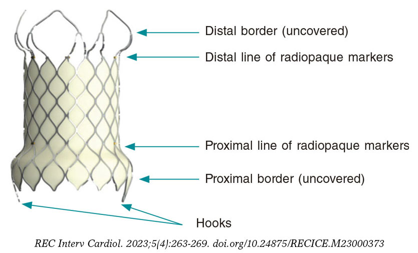
Eighty-seven procedures were reported, of which 51 (58.6%) were performed in patients older than 18 years. The hybrid approach was used in 2 cases, while the fully percutaneous approach was used in the remaining cases. The pulmonary position was predominant (96.5%), with 2 successful valve implantations being performed in the tricuspid position and 1 in the mitral position. The anatomical substrate of implantation in the pulmonary position had the following distribution: 33 in the surgical conduit, 31 in the native tract, followed by 20 valve-in-valve procedures.
Complications
Morbidity and mortality data were reported for 2401 interventional procedures, with 35 serious adverse events (table 4), including 6 deaths, which translated into a rate of major complication of 1.4% and a mortality rate of 0.2%. The categories associated with higher morbidity rates were percutaneous valve implantation (8%), other angioplasty procedures (6%), and VSD closure (5.2%). The most common complications were device embolizations (8 cases): 4 in ASD closures, 2 in patent ductus arteriosus closures, and 2 stents implanted in the setting of pulmonary angioplasty procedures; surgical removal of the embolized valve was required in only 1 case of ASD closure. Less frequent were vascular complications (6 cases), 3 of them being associated with pulmonary angioplasty procedures. There were 4 cases of severe arrhythmias, including 2 cases of cardiac arrest requiring bailout extracorporeal membrane oxygenation.
Table 4. Distribution of major complications and reported deaths in various interventional procedures
| Procedure | n | Major complications | Deaths |
|---|---|---|---|
| Congenital aortic valvuloplasty | 67 | 3 (6.5) – 1 severe aortic regurgitation – 1 unspecified – 1 death |
1 |
| Valvuloplastia pulmonar congénita | 138a (111) | 2 (1.8) – 1 tricuspid valve rupture – 1 unspecified |
0 |
| Valvuloplastia mitral congénita | 0 | 0 | 0 |
| Angioplastia pulmonar | 135b (102) | 1 (0.9) – 1 unspecified |
0 |
| Angioplastia ramas pulmonares | 227c (202) | 6 (2.9) – 3 vascular dissections – 1 pulmonary hemorrhage – 2 stent embolizations |
0 |
| Angioplastia aórtica | 124d (102) | 3 (2.9) – 2 vascular dissections – 1 death |
1 |
| Otras angioplastias | 100 | 6 (6) – 1 coronary thrombosis – 1 CPR-ECMO – 1 vascular dissection – 1 neurological event – 2 deaths |
2 |
| Cierre de comunicación interauricular/foramen oval | 1135 | 5 (0.4) – 4 embolizations (1 required surgery) – 1 neurological event |
0 |
| Cierre de conducto | 262 | 3 (1.1) – 2 embolizations not requiring surgery – 1 death |
1 |
| Cierre de comunicación interventricular | 38 | 2 (5.2) – 1 atrioventricular block – 1 CPR-ECMO |
0 |
| Otras oclusiones | 91 | 0 | 0 |
| Retirada de cuerpo extraño | 23 | 0 | 0 |
| Atrioseptostomía | 72 | 1 (1.3) – 1 unspecified |
0 |
| Implantación de válvula percutánea | 87 | 4 (8.0) – 1 vascular dissection – 1 pulmonary duct dissection – 1 ventricular tachycardia – 1 death |
1 |
| Total | 2508e (2401) | 35 (1.4) | 6 (0.2) |
|
CPR, cardiopulmonary resuscitation; ECMO, extracorporeal membrane oxygenation. a Percentages calculated based on 111 reported cases. b Percentages calculated based on 102 reported cases. c Percentages calculated based on 202 reported cases. d Percentages calculated based on 102 reported cases. e Percentages calculated based on 2411 reported cases. Data are expressed as n (%). |
|||
DISCUSSION
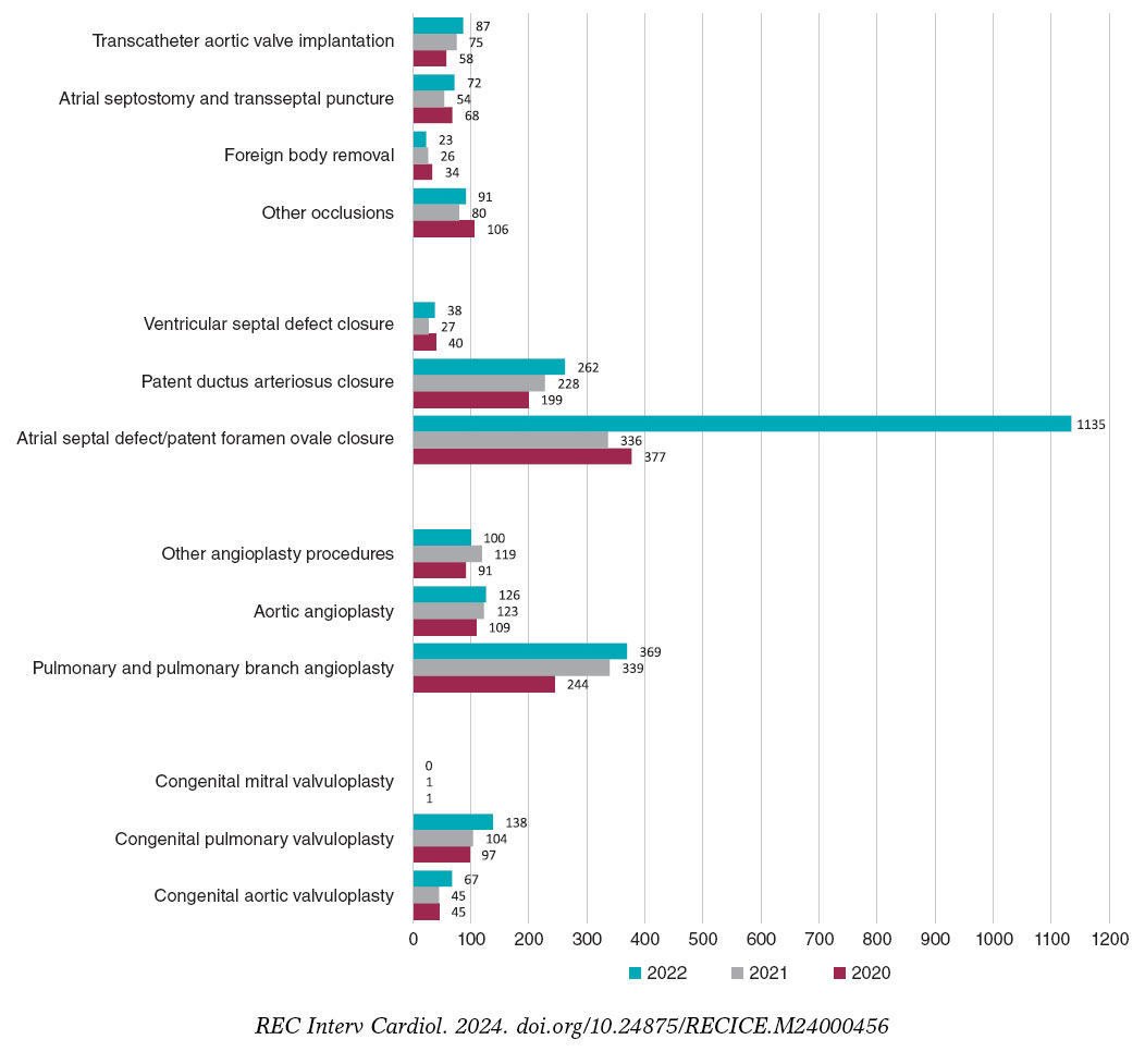
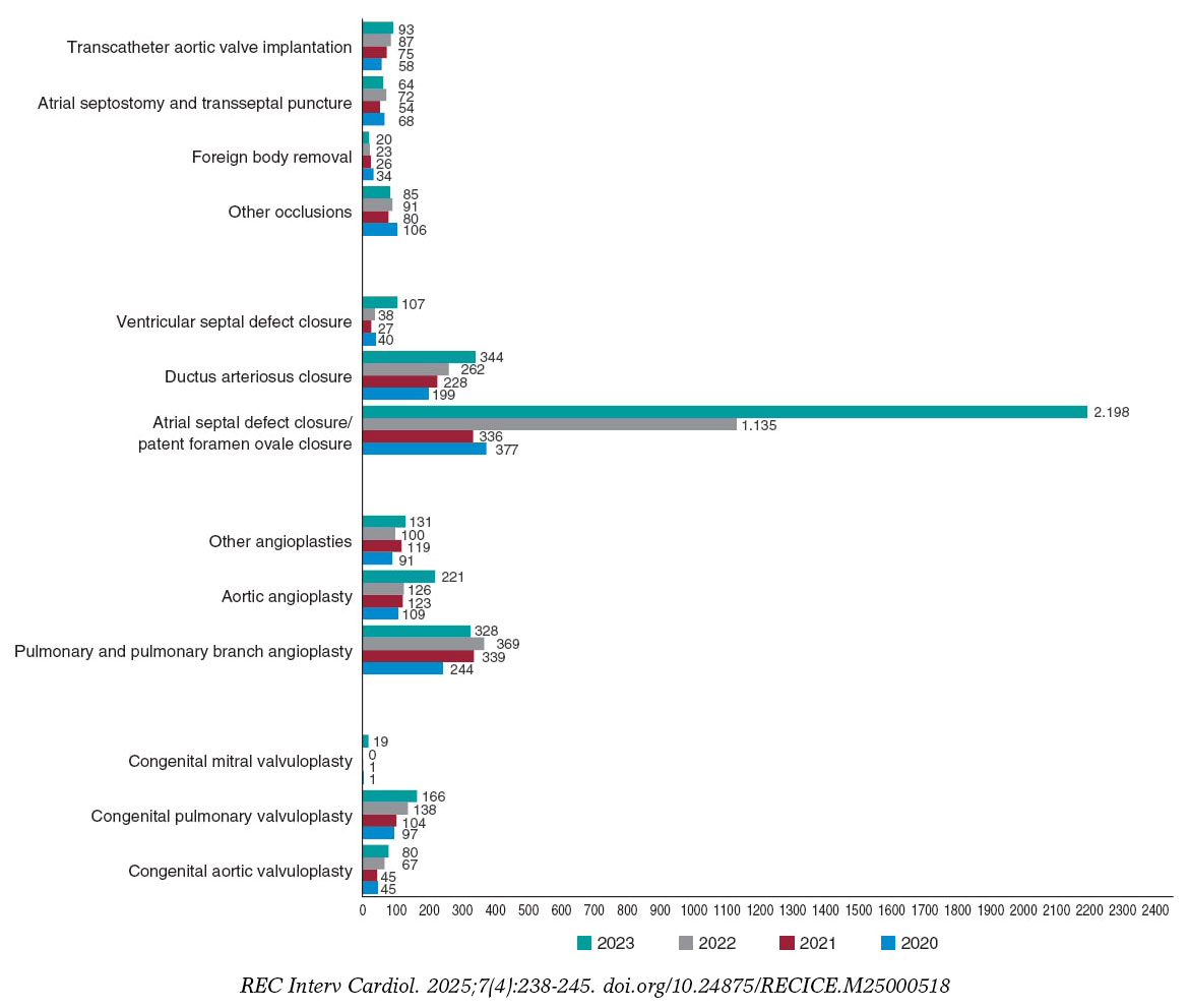
To date, one of the main weaknesses of this registry has been its limitations in adequately assessing the interventional activity carried out in the context of adult congenital heart disease. For this reason, and as the most significant novel addition to this report, we included data from 99 hospitals reporting their activities in adult congenital heart disease to the 2022 Spanish Registry of Cardiac Catheterization and Interventional Cardiology in the analysis of the various interventional categories. This has resulted in a significant increase in catheterization volume, totaling 3649 procedures (1002 more than in 2021). Their comparison with the activity conducted in previous years and the significant increase in registered procedures should be analyzed considering this methodological difference, and taking into account the increase in participating centers, 6 more than in 2021 (figure 1).
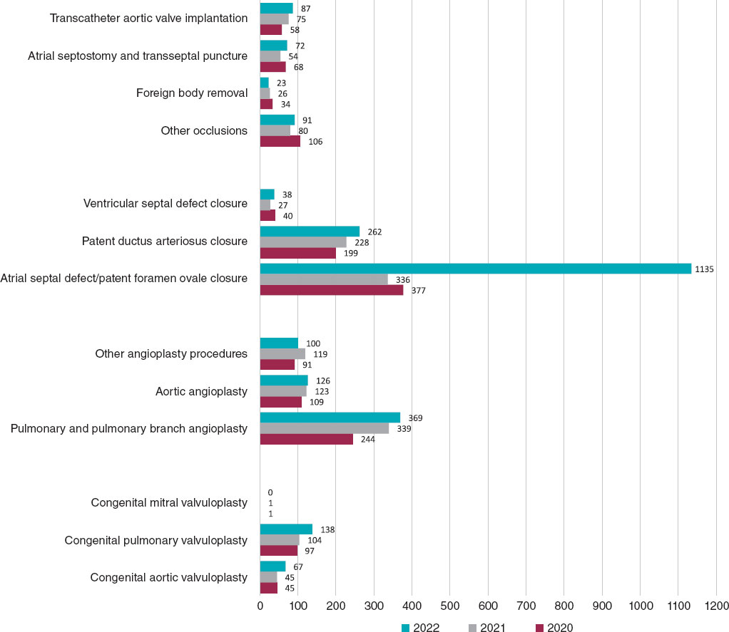
Figure 1. Comparison of the number of interventional procedures performed in 2020, 2021, and 2022.
The total number of registered interventional procedures was 2508, with notable increases in techniques such as ASD closure, aortic and pulmonary valvuloplasty, atrial septostomy, and VSD closure. A total of 55% of cardiac catheterizations were performed in patients older than 18 years (compared with 32% in 2021), demonstrating an improvement in the representation of interventional procedures for adult congenital heart diseases. Once again, in the pediatric setting, we noted that fetal interventional activity in Spain is very limited, with only 3 reported cases (2 aortic valvuloplasty procedures and 1 pulmonary valvuloplasty procedure), despite evidence of its value and effectiveness in these and other prenatal scenarios, such as pulmonary atresia with intact ventricular septum and hypoplastic left heart syndrome.7
The reported data on the effectiveness of various interventional techniques yielded an overall success rate of 97.6% (compared with 95% in 2021) and a mortality rate of 0.2% (the same as in 2021), with 6 procedure-related deaths. These results are consistent with those reported from most international studies to date.8,9 The rate of serious adverse events of 1.4% is the lowest reported so far (2% in 2020 and 2.7% in 2022), with a decrease in the frequency of all types of complications reported. Device embolizations continue to account for the highest number of cases, amounting to 22.5% overall, followed by vascular complications (20% overall).
The volume of valvuloplasty procedures has significantly increased with respect to 2021: a 48.9% increase in aortic valvuloplasty and a 32.7% increase in pulmonary valvuloplasty. For the first time, most cases involving one of these 2 techniques involved patients older than 1 year. In aortic valvuloplasty, the rate of serious events (6.5%) decreased compared with the previous year (11.1%), although with 1 associated death. The report shows that pulmonary valvuloplasty has become established as one of the techniques with the best results, with a 99.2% efficacy rate and a 1.8% complication rate. These data support the value of pulmonary valvuloplasty as the technique of choice in congenital pulmonary valve stenosis in our setting. However, its mid- and long-term outcomes may be influenced by unspecified anatomical and genetic factors.10
Both in pulmonary angioplasty procedures (of native tract or ducts) and pulmonary branch angioplasty procedures, stent implantation has surpassed conventional balloon dilation as the technique of choice, which has again reduced the use of cutting balloons. The most widely performed aortic angioplasty procedures continue to be aortic arch and isthmus dilatation, which are performed in almost all patients; of note, in this context, the increase in covered stent implantation, which, for the first time, has surpassed other dilation techniques. This increase could be explained by the intention to improve the safety of the procedure by reducing damage to the aortic wall in certain scenarios.11 Furthermore, the availability of covered stents with lower implantation profiles has facilitated their use in pediatric patients of increasingly lower weight and younger age.12
ASD closure remained the most widely performed interventional technique in the registry (45.2% of all interventional catheterizations). The inclusion of patent foramen ovale closure as a procedure within this category and its classification as a congenital heart disease may be controversial but can be reevaluated in future reports. Its rarity in the pediatric setting contrasts with its increasing application in adults, confirming the maturity of the technique and the widespread acceptance of the scientific evidence supporting its use.13 Transesophageal echocardiography guidance remains the usual imaging modality for ASD closure; both intracardiac echocardiography and balloon sizing of the defect are infrequent.
A notable finding was the increasing use of patent ductus arteriosus closure in the group of premature newborns (9.4% overall), as well as confirmation of the preference for the transcatheter option over surgery for these pediatric patients in our setting.14 Antegrade venous access and the use of occlusive devices remain widespread procedures in a consolidated technique that has one of the best effectiveness rates in the registry (98.9%).
The reported data on the safety and efficacy of VSD closure show substantial improvement compared with previous reports: the major complication rate decreased from 18% in 2021 to 5.2% in 2022, while the success rate increased from 77.3% in 2021 to 96.7% in 2022. These figures reflect a change in trend, which could be related to the introduction of new closure devices, and the adoption of technical changes facilitating their approach.15-17 All of this would facilitate the widespread use of the procedure, whose frequency has increased significantly by up to 40.7% compared with the previous year. The increase in the number of cases registered in patients older than 18 years was notable, reaching 38% overall (compared with 22% in 2021).
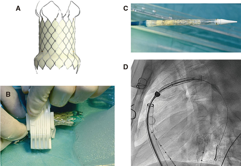
A 16% volume increase and a significant improvement in the reported safety and efficacy data of transcatheter aortic valve implantation were also reported, of which approximately 60% were performed in patients older than 18 years. There was a decrease in the tricuspid position as the anatomical substrate for implantation (from 10 cases in 2021 down to only 2 cases in 2022), at a time when transcatheter aortic valve implantation has reached an unprecedented growth as a structural heart procedure in Spain.5 Access to new valves—especially self-expanding valves—and the continuous publication of scientific evidence endorsing the results of this technique, continue to enhance the expectations of the percutaneous management of patients with right ventricular outflow tract dysfunction in all anatomical scenarios.18,19
Limitations
The characteristics of this registry may be weakened by its retrospective, voluntary, and unaudited design. Expanding the collected data on certain techniques of special interest would help improve its quality and should be considered in future reports.
CONCLUSIONS
The main finding of this report is the significant increase in the number of interventional procedures recorded compared with previous years, which was closely related to the increase in participating centers. There has been significant growth in aortic valvuloplasty, ASD closure, and VSD closure procedures. The data obtained provide a realistic overview of interventional activity in congenital heart diseases in Spain among all age groups. The reported safety and efficacy results demonstrate the consolidation of most techniques in our setting and are consistent with those published in other international studies.
The incorporation of a greater number of centers with interventional activity in congenital heart diseases into the registry will optimize the quality and reliability of the information generated.
FUNDING
None declared.
ETHICAL CONSIDERATIONS
Due to the methodological characteristics of the study and its nature as solely an activity registry, there was no requirement for approval from ethics committees or signing of informed consent forms.
The characteristics of the present work exclude the consideration of possible variables of sex and gender.
STATEMENT ON THE USE OF ARTIFICIAL INTELLIGENCE
No artificial intelligence tools were used in the preparation of this article.
AUTHORS’ CONTRIBUTIONS
All authors contributed substantially to data collection and the critical review of this work. F. Ballesteros Tejerizo and F. Coserría Sánchez wrote the article.
CONFLICTS OF INTEREST
S. Ojeda Pineda is an associate editor of REC: Interventional Cardiology; the journal’s editorial procedure to ensure the impartial processing of the manuscript has been followed. The remaining authors declare no conflicts of interest.
WHAT IS KNOWN ABOUT THE TOPIC?
- Cardiac catheterization remains an indispensable procedure in the management of patients with congenital heart diseases.
- The existence of a national registry of pediatric percutaneous procedures and adult congenital heart diseases is essential to understand the current panorama of interventional cardiology in Spain and generate valuable information for professionals, patients, and families.
- The continuity of this registry allows understanding the level of implementation and results of various techniques, as well as their variation over time.
WHAT DOES THIS STUDY ADD?
- Some methodological changes and the gradual increase in the number of centers participating in the registry have enabled the collection of more realistic information on interventional activity for congenital heart diseases among all age groups in Spain.
- A highly significant increase in interventional procedures performed in 2022 was reported, with ASD and VSD closure and aortic valvuloplasty procedures being the techniques experiencing the greatest growth.
- The most widely performed procedures continue to be ASD closure, patent ductus arteriosus closure, and pulmonary artery branch angioplasty.
- The most frequent procedure-related adverse events were device embolizations and vascular complications.
REFERENCES
1. Ballesteros Tejerizo F, Coserría Sánchez F, Romaguera R, et al. Spanish Cardiac Catheterization in Congenital Heart Diseases Registry. First Official Report from ACI-SEC and GTH-SECPCC (2020). REC Interv Cardiol. 2022;4:173-180.
2. Ballesteros Tejerizo F, Coserría Sánchez F, Freixa X, et al. Spanish cardiac catheterization in congenital heart diseases registry. Second official report from the ACI-SEC and the GTH-SECPCC (2021). REC Interv Cardiol. 2023;5:185-192.
3. Romaguera R, Ojeda S, Cruz-González I, et al. Spanish Cardiac Catheterization and Coronary Intervention Registry. 30th Official Report of the Interventional Cardiology Association of the Spanish Society of Cardiology (1990-2020) in the year of the COVID-19 pandemic. Rev Esp Cardiol. 2021;74:1096-1106.
4. Freixa X, Jurado-Roman A, Cid B, et al. Spanish cardiac catheterization and coronary intervention registry. 31st Official Report of the Interventional Cardiology Association of the Spanish Society of Cardiology (1990-2021). Rev Esp Cardiol. 2022;75:1040-1049.
5. Jurado-Román A, Freixa X, Cid B, et al. Spanish cardiac catheterization and coronary intervention registry. 32nd Official Report of the Interventional Cardiology Association of the Spanish Society of Cardiology (1990-2022). Rev Esp Cardiol. 2023;76:1021-1031.
6. Asociación de Cardiología Intervencionista de la Sociedad Española de Cardiología. Registro de Actividad ACI-SEC. Available at:http://www.registroactividadacisec.es. Accessed 21 Jun 2023.
7. Friedman KG, Tworetzky W. Fetal cardiac interventions:Where do we stand?Arch Cardiovasc Dis. 2020;113:121-128.
8. Kevin D, Wei Du, Fleming GA, et al. Validation and refinement of the catheterization RISK score for pediatrics (CRISP score):An analysis from the congenital cardiac interventional study consortium. Catheter Cardiovasc Interv. 2019;93:97-104.
9. Quinn BP, Ye M, Gauvreau K, et al. Procedural Risk in Congenital Cardiac Catheterization (PREDIC3T). J Am Heart Assoc. 2022;11:022832.
10. Hansen RL, Naimi I, Wang H, et al. Long-term outcomes up to 25 years following balloon pulmonary valvuloplasty:a multicenter study. Congenit Heart Dis. 2019;14:1037-1045.
11. Stassen J, De Meester P, Troost E, et al. Covered stent placement for treatment of coarctation of the aorta:immediate and long-term results. Acta Cardiol. 2021;76:464-472.
12. Al Balushi A, Pascall E, Jones MI, Qureshi S, Butera G. Initial experience with a novel ePTFE-covered balloon expandable stent in patients with near-atretic or severe aortic coarctation and small femoral arterial access. Cardiol Young. 2021;31:224-228.
13. Saver JL, Carroll JD, Thaler DE, et al.;RESPECT Investigators. Long-Term Outcomes of Patent Foramen Ovale Closure or Medical Therapy after Stroke. N Engl J Med. 2017;377:1022-1032.
14. Rodríguez-Ogando A, Ballesteros Tejerizo F, Blanco Bravo D, et al. Transcatheter Occlusion of Patent Ductus Arteriosus in Preterm Infants Weighing Less Than 2 kg With the Amplatzer Duct Occluder II Additional Sizes Device. Rev Esp Cardiol. 2018;71:861-876.
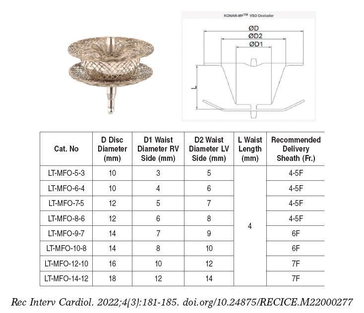
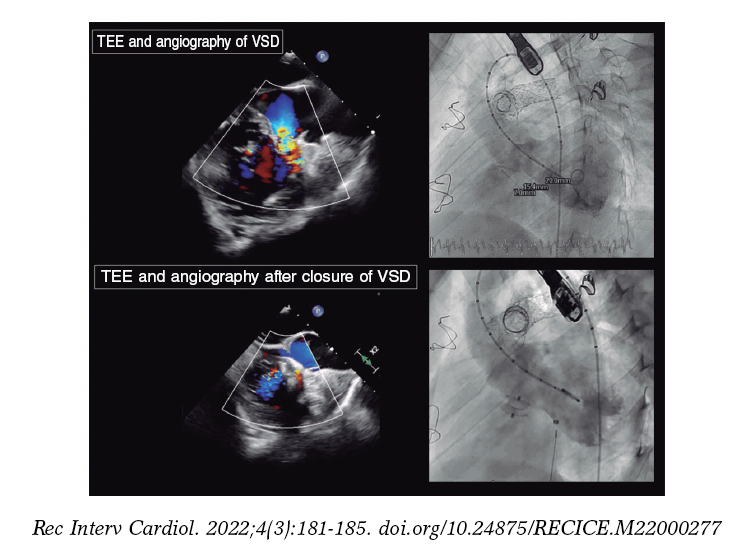
15. Alvarez-Fuente M, Carrasco JI, Insa B, et al. Percutaneous closure of ventricular septal defect with the KONAR-MF device. REC Interv Cardiol. 2022;4:181-185.
16. Rasines Rodri?guez A, Aristoy Zabaleta MM, Abelleira Pardeiro, et al. Retrograde closure of perimembranous ventricular septal defects. A paradigm shift. REC Interv Cardiol. 2023;5:73-75.
17. Nistor IA, Mesa Rubio D, Pan Álvarez-Ossorio M. Percutaneous VSD closure with the KONAR-MF occluder:fusion helps. Rev Esp Cardiol. 2023;77:106.
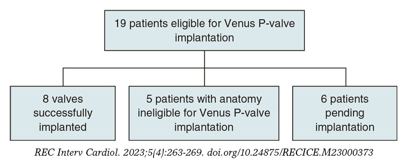
18. A?lvarez-Fuente M, Toledano M, Hernández I, et al. Initial experience with the new percutaneous pulmonary self-expandable Venus P-valve. REC Interv Cardiol. 2023;5:263-269.
19. Hascoët S, Bentham JR, Giugno L, et al. Outcomes of transcatheter pulmonary SAPIEN 3 implantation:an international registry. Eur Heart J. 2024;45:198-210.
* Corresponding author.
E-mail address: fernandoballe@gmail.com (F. Ballesteros Tejerizo).
ABSTRACT
Introduction and objectives: Percutaneous pulmonary valve implantation is currently a common procedure in patients with congenital heart disease with a dysfunctional right ventricular outflow tract. Until April 2022, there were only balloon-expandable valves available in Europe, which did not cover the needs of the different anatomies of the right ventricular outflow tract. Since that date we have available the self-expandible Venus P-valve (Venus MedTech, China). We present the initial experience with this new percutaneous pulmonary valve in our center.
Methods: Description of the valve implants with the new self-expandible valve performed between September and November 2022.
Results: Eight valve implants have been performed, all successful and without severe complications during the procedure. All patients had severe pulmonary regurgitation with a dilated right ventricle and severe dilatation of the pulmonary trunk and were not good candidates for percutaneous balloon-expandable valves. Five patients had a tetralogy of Fallot. In 7 patients, the implant was performed through the femoral vein and in one through jugular access. As a safety measure, all valves were implanted through a DrySeal sheath (Gore, W.L. Gore & Associates, Inc., United States). The mean hospital stay was 3-day.
Conclusions: Valve implantation with the new self-expandible Venus P-valve was, in our preliminary experience, a safe and feasible procedure, allowing us to treat very dilated right outflow tracts, not suitable for the current balloon-expandable valves.
Keywords: Percutaneous valve implantation. Venus P-valve. Tetralogy of Fallot. Pulmonary regurgitation. Pulmonary valve. Congenital heart disease.
RESUMEN
Introducción y objetivos: El implante percutÁneo de vÁlvula pulmonar es, actualmente, un procedimiento habitual en pacientes con cardiopatías congénitas con un tracto de salida del ventrículo derecho disfuncionante. Hasta abril de 2022, en Europa solo estaban disponibles las vÁlvulas expandibles con balón, que no cubrían las necesidades de las distintas anatomías del tracto de salida derecho. Desde esa fecha estÁ disponible la vÁlvula autoexpandible Venus P ( Venus MedTech, China). Presentamos la experiencia inicial en nuestro centro con esta nueva vÁlvula pulmonar para implante percutÁneo.
Métodos: Descripción de los implantes valvulares con la nueva vÁlvula autoexpandible realizados entre septiembre y noviembre de 2022.
Resultados: Se han realizado 8 implantes valvulares, todos con éxito y sin complicaciones graves durante el procedimiento. Todos los pacientes presentaban insuficiencia pulmonar grave con repercusión sobre el ventrículo derecho y dilatación del tronco pulmonar, y no eran buenos candidatos para las vÁlvulas expandibles con balón. Cinco pacientes tenían una tetralogía de Fallot de base. En 7 pacientes el implante se llevó a cabo por vía femoral y en 1 por vía yugular. Como medida de seguridad, en todos los pacientes el implante se hizo a través de una vaina DrySeal (Gore, W.L. Gore & Associates, Inc., Estados Unidos). La media de tiempo de ingreso fue de 3 días.
Conclusiones: El implante de la nueva vÁlvula autoexpandible Venus P fue, en nuestra experiencia preliminar, un procedimiento seguro y factible, que permite valvular tractos de salida derechos muy dilatados con contraindicación para las actuales vÁlvulas expandibles con balón.
Palabras clave: Implante percutÁneo valvular. VÁlvula pulmonar Venus P. Tetralogía de Fallot. Insuficiencia pulmonar. VÁlvula pulmonar. Cardiopatías congénitas.
Abbreviations
CT: computed tomography. LMCA: left main coronary artery. MRI: magnetic resonance imaging. PAT: pulmonary arterial trunk. RV: right ventricle. RVOT: right ventricular outflow tract.
INTRODUCTION
Currently, transcatheter pulmonary valve implantation is a common procedure in patients with congenital heart disease and dysfunctional right ventricular outflow tract (RVOT).1 Transcatheter balloon-expandable pulmonary valves (Melody by Medtronic Inc., United States, and Sapien by Edwards Lifescience, United States, both with CE marking) have an indication for conduit, native tract, and prosthetic valve implantation.1,3 However, a large number of patients—most with tetralogy of Fallot—who require pulmonary valve implantation are treated with transannular repair or post-commissurotomy pulmonary regurgitation or valvuloplasty for pulmonary valve stenosis. These patients have very pulsatile outflow tracts with larger sizes compared to the ones of current balloon-expandable valves (22 mm and 29 mm for Melody and Edwards, respectively).4 Not even the 32 mm Myval device (Meril Life Sciences Pvt. Ltd., India) without an indication for pulmonary valve implantation would be adequate for the largest RVOTs out there.
Back in April 2022, the self-expanding Venus P-valve (Venus MedTech, China) achieved the CE marking, and became an actual alternative for the largest native tracts.
This is our initial experience with this new transcatheter pulmonary valve at our center and in our country.
METHODS
Valve description
The structure of the Venus-P valve consists of a nitinol stent. Both the leaflets, and the stent coverage are made of porcine pericardium. Nitinol provides the stent with some sort of shape memory so it can adapt to the pulmonary arterial trunk (PAT) without compressing neighboring structures. This valve is available in sizes from 28 mm to 36 mm in diameter with 2 mm increases (figure 1). It can be used with the largest caliber native tracts.5 The Venus P-valve stent has a diabolo-shaped configuration and adds 10 mm to the borders of the central region (figure 1). It is wider in its borders because it has been designed for tubular PAT implantation without distal or pulmonary artery stenosis. Both the central region and the proximal border are covered with porcine pericardium to prevent paravalvular leak. The distal border remains uncovered to avoid occluding the pulmonary arteries (figure 2).6 The stent has radiopaque marks in the distal border of its tubular region and in its proximal border both indicative of the degree of the porcine valve implantation. The valve crimping system on its delivery system is performed under ice water. In these conditions, nitinol becomes softer and can be crimped onto the delivery system (figure 3). The valve is then fixed to the delivery system through 2 small hooks (figure 2). Once the valve has been attached and its size reduced, it is covered with the delivery sheath capsule in such a way that the valve enters the patient fully covered (figure 3 and figure 4) [22-Fr sheaths in 28 mm and 30 mm valves, and 24-Fr sheaths in the largest ones (> 30 mm)]. Once in the pulmonary tree and in the desired location, the delivery sheath capsule will be retracted so the valve can regain its diabolo-shaped configuration when entering blood flow at 36 ºC to 37 °C temperature.
Figure 1. Selection of different valvular sizes with the dimensions of the different parts of the valve. RV, right ventricle.
Figure 2. Image of the Venus P-valve with its typical diabolo-shaped configuration, and coverage of the entire valve leaving both the distal border and the radiopaque markers uncovered.
Figure 3. A: Venus P-valve prior to crimping. B: valve crimping onto the delivery system inside a frozen physiological saline solution with a specific crimping system. C: crimped valve inside the delivery system while covered by a capsule. D: fluoroscopy showing the valve in the delivery system inside the capsule already inserted into the patient’s pulmonary artery.
Procedural description
Cardiac catheterization was performed under general anesthesia while the patient remained intubated and with heparinization at 100 U/kg. Two femoral veins where cannulated, 1 for diagnostic catheterization, cutting, and advance of the valve delivery system, and the other one to perform the follow-up angiography with a pigtail catheter in the RVOT during valve implantation (figure 4). The coronary arteries of all the patients were interrogated through aortograms or selective coronary angiographies with the same 34 mm cutting balloon (Sizing Balloon, AGA Medical Corp., United States) inflated in the RVOT to discard the risk of coronary compression during the procedure (figure 5B). Similarly, the RVOT was cut with the same 34 mm balloon to see if the anatomy was viable and choose the right valve diameter and length. Cutting was considered occlusive when aortic pressure dropped during balloon inflation and the lack of right ventricular (RV) flow to the pulmonary arteries was angiographically confirmed through an intracoronary injection of contrast into the RV during the balloon peak inflation rate (figure 5A).
Figure 4. Valve implantation process. A-D: slow capsule removal so the valve can adapt to the left ventricular outflow tract (LVOT). Angiography monitorization with the pigtail catheter placed in the LVOT. E: final outcomes after valve implantation.
Figure 5. Right ventricular outflow tract cutting with a 34 mm cutting balloon. A: simultaneous angiography in the right ventricle showing the complete occlusion of the RVOT by the balloon that shows a 27 mm notch at stenosis level. B: coronary artery interrogation during cutting balloon inflation without coronary compromise.
Although the imaging modalities performed while planning (computed tomography scan [CT] with or without magnetic resonance imaging (MRI)] inform us on the most appropriate valvular size for each patient, size was picked based on angiography measurements (30° lateral and 30°cranial right anterior oblique), and on the diameter and location of the notch seen in the balloon during occlusive cutting. Balloon cutting allows us to assess the compliance of the PAT, something that is merely suggested on the MRI. Therefore, this is an essential step that should be made before selecting the size of the valve. We select a 2 mm-to-4 mm larger valve compared to the waist of the cutting balloon with a length that should leave the distal border at pulmonary bifurcation level, and the diabolo proximal border at RV level.
The Venus P-valve is implanted without the need for previous stenting to create a landing zone. A Lunderquist high-support guidewire is placed (Cook Medical, Denmark) preferably in the left pulmonary artery. In the presence of stenosis, hypoplasia or unfavorable angle, the guidewire is placed in the right pulmonary artery. The delivery system is advanced through a 65 cm 26-Fr or 24-Fr Dryseal sheath (Gore, W.L. Gore & Associates, Inc., United States) for valves > 30 mm or < 30 mm in diameter, respectively. Once the Dryseal sheath is in position into the selected pulmonary artery, the Venus P-valve delivery system is advanced. Afterwards, the Dryseal sheath is retracted and the correct position of the capsule into the pulmonary artery is verified. Then, the valve is slowly uncovered by withdrawing the capsule (with a clockwise twist of the wheel of the delivery system). As it gradually enters the patient’s bloodstream, the valve regains its normal diabolo-shaped configuration. When its most distal border has been partially adapted to the pulmonary branch, the whole system is then smoothly removed to the PAT and the valve is slowly uncovered while checking—through pigtail catheter injections into the RVOT—that the valve is in position (figure 4). The valve has 2 lines of radiopaque markers to guide implantation. The distal marks that indicate the distal border of the stent tubular region will remain at bifurcation level. The proximal ones should remain at PAT narrowest region or native annulus level (in the location of the notch seen during balloon cutting), thus reducing the risk of embolization. The valve proximal region—10 mm larger than the central one—will remain, in most cases, in the infundibulum of the RV.
Procedure should be performed through a Dryseal sheath to facilitate the maneuverability of the valve delivery system and guarantee safe valve recapture should repositioning be required. The valve can be recaptured until half of its structure has been uncovered.
An aortogram was performed at the end of all procedures (preferably 30° caudal and 20º left anterior oblique) to confirm the lack of coronary artery compression (figure 6).
Figure 6. Final aortogram for coronary artery assessment. Arrows are indicative of the trajectory of the left anterior descending coronary artery.
Patient selection
A previous study through diagnostic catheterization or MRI with or without CT scan was conducted to see whether the anatomy of potentially eligible patients with valve implantation criteria according to the clinical practice guidelines published by the ESC7,8 was ripe for Venus P-valve implantation (figure 7). CT or MRI acquired images were analyzed by Venus Medtech image technicians who video-called our hospital heart team to discuss the convenience of valve implantation and proper valvular size that best suited the patient’s RVOT size. However, the final size was not decided until balloon cutting was used during cardiac catheterization.
Figure 7. Flowchart of patients included in the study.
This first imaging study discarded 5 patients with unsuitable anatomies for this kind of valve: 2 patients with stents and pulmonary artery stenoses, 2 patients with larger RVOTs compared to the sizes recommended for this kind of valve, and 1 patient with pyramid-shaped right ventricular outflow tract.
Study description
This was a prospective study of the first patients treated with Venus P-valve implantation conducted at our center from September 20th through November 4th, 2022. These are the very first implantations of this type of valve ever performed in our country.
Inclusion criteria
This study included patients with dysfunctional native RVOTs and an indication for pulmonary valve implantation in whom diagnostic catheterization and cutting test allowed such procedure.
Variables
Demographic and anthropometric data were collected, as well as imaging modality and procedural data to conduct a descriptive analysis of our own experience.
Definitions
Complications were categorized as minor or major. The later were death, potentially life-threatening adverse events, and events requiring surgery (embolization, myocardial perforation, vascular rupture, residual PR, hemolysis, valvular lesion). The former were complications that resolve spontaneously or subside with clinical treatment without potentially fatal outcomes (vascular access problems, fever, neuroapraxias, etc.). Implantation was considered successful in the absence of major complications 24 hours after the procedure.
RESULTS
A total of 8 Venus P-valves were implanted in 8 patients at our center from September 20th through November 4th, 2022. The underlying conditions were tetralogy of Fallot (5 patients), pulmonary atresia with ventricular septal defect (1 patient), atrial septal defect with pulmonary stenosis (1 patient), and ventricular septal defect and pulmonary artery banding (1 patient) who required RVOT dilatation in its configuration. All the patients had a transannular patch. Also, all patients had severe pulmonary regurgitation with RV repercussion and PAT dilatation, and were ineligible for transcatheter balloon-expandable valve implantation due to the size of their RVOTs. table 1 shows a overall description of the patients.
Table 1. Overall description of the sample
| Patient | Sex | Age (years) |
Weight (kg) |
RV (mlLm2) |
PAT (mm) MRI |
PAT (mm) CT |
PAT (mm) angiography |
Cutting balloon (mm) |
Valvular size |
|---|---|---|---|---|---|---|---|---|---|
| 1 | W | 41 | 56 | 122 | 28 | NA | 26 | 26 | 30-25 |
| 2 | M | 34 | 62 | 164 | 28 | 27 | 28 | 28 | 32-25 |
| 3 | W | 25 | 66 | NA | NA | NA | 28 | 28 | 32-25 |
| 4 | M | 33 | 90 | NA | NA | 33 | 31 | 32 | 36-25 |
| 5 | M | 34 | 68 | 134 | 31 | NA | 26 | 27 | 34-25 |
| 6 | M | 17 | 63 | 173 | 29 | 31 | 31 | 31 | 34-25 |
| 7 | M | 45 | 68 | NA | NA | 34 | 26 | 32 | 34-30 |
| 8 | W | 43 | 42 | 130 | 25 | NA | 24 | 24 | 28-25 |
|
CT, computed tomography scan; M, man; MRI, magnetic resonance imaging; NA, not available; PAT, pulmonary arterial trunk; RV, right ventricle; W, woman. |
|||||||||
All patients had dilated PATs as seen on the CT scan or MRI—meaning they were suboptimal candidates for balloon-expandable valve implantation—and tubular PATs without stenoses in, at least, 1 pulmonary artery.
The valves were successfully and uneventfully implanted in all the patients with optimal valvular competence immediately after implantation. A total of 7 implantations were performed via femoral access, and 1 via right jugular vein due to bilateral femoral venous thrombosis. High-support guidewires were placed in the left pulmonary artery (6 cases), and right pulmonary artery (2 cases, 1 due to moderate left pulmonary artery stenosis, and the other one because it was a jugular access). In 7 and 1 cases, respectively, 26-Fr and 24-Fr Dryseal sheaths were used through which implantation occurred. The length of the valves implanted was 25 mm (n = 7) and 30 mm (n = 1).
The median fluoroscopy time was 34 min (interquartile range, 32-37), and the mean radiation dose, 307 mGy/m2 (standard deviation, 64.4). We saw an adequate correlation between the CT and MRI measurements of the PAT and the angiography measurements and the cutting balloon.
No severe complications were reported in any of the cases. In 1 case, while the valve was being deployed, 1 of the proximal hooks got trapped in the delivery system due to incomplete removal of the capsule covering the valve. It, however, resolved uneventfully (figure 8). A total of 3 patients had mild thoracic pain 24 hours after implantation not showing elevated troponin levels or ECG abnormalities. Also, 1 patient complained of right scapula pain. One of the patients had common ventricular extrasystole that started right after valve implantation and subsided spontaneously within the next 48 hours not requiring any therapy at discharge. One patient broke a fever 48 hours after implantation without elevation of acute phase reactants.
Figure 8. Arrows indicate, in 2 different projections, that 1 of the attachment hooks is still trapped in the delivery system preventing full valve deployment.
The mean length of stay was 3 days (range 2-4). All patients were discharged on acetylsalicylic acid.
DISCUSSION
Transcatheter pulmonary valve implantation is, currently, a procedure widely performed in the cath labs of congenital coronary care units. Up until now, the valves available—with sizes up to 29 mm—left a significant number of patients without transcatheter therapeutic options available. In some cases, off-label techniques were used (several stents implanted in the PAT to reduce its caliber, stent landing in the left pulmonary artery, etc.) or larger sized balloon-expandable valve implantation with an indication for the aorta (32 mm Myval valve), but not approved for the RVOT.9
Among the main advantages of the Venus P-valve is the possibility or performing one-stage diagnostic catheterizations and valve implantations since previous stenting is not required to create a landing zone (as it was the case with the Melody and Edwards valves in several native tracts). In the most dilated pulmonary arterial trunks (> 24 mm-to-26 mm) one-stage stenting is the gold standard. Then, wait for, at least, 6 weeks until endothelization occurs to minimize the risk of embolization while the valve is being implanted, which requires a second catheterization to implant the valve. Another advantage of the Venus P-valve is how easy it is to use. It is a short procedure with 34 min fluoroscopy times in our cases (shorter to the times reported in our series of native RVOTs and other valves between 40 min and 50 min).
The flexibility of the valve allows PAT adaptation without exerting radial strength on adjacent structures, which is a plus for cases of coronary RVOT-related anatomies.
The short 25 mm valve was used in 7 cases since these patients’ PAT anatomy allowed such length. With longer length, less flexibility for the valve and higher risk of long-term fractures. Up until today, a rate of fractures between 11% and 27% has been reported without associated complications or loss of valvular competence associated with the fractures.5,10
We should pay special attention to the complete removal of the capsule at the end of valve deployment because, if removal is incomplete as it happened with our case (figure 8), hooks can’t be released, and the valve does not get deployed with the corresponding risk of RV embolization.11 To make sure that the system has been released, the hooks should be checked in, at least, 2 different views or projections.6
The diabolo-shaped configuration makes this valve unsuitable for all RVOT anatomies. In cases of distal stenosis at PAT level or at the origin of pulmonary arteries this valve is ill-advised because such stenoses would be limiting the opening of the valve distal border. Regarding this shape—wider in its borders—the onset of transient ventricular extrasystole due to contact with the infundibulum proximal border has been reported. However, to this date, no arrhythmias have ever been reported requiring ablation or cardioversion. In our own experience, only 1 patient had frequent extrasystoles, yet no sustained ventricular arrhythmias were ever reported.
The clinical trial (NCT 02846753) conducted to obtain the CE marking revealed the presence of isolated endocarditis (incidence rate of 1.2%). When long-term follow-up results become available, the actual rates of endocarditis, fractures, and valvular dysfunction will be assessed.
Limitations
The short follow-up of patients is this study main limitation. We’ve been closely monitoring these patients including the use of Holter monitor and imaging modalities (echocardiography, CT scan, and MRI) to properly assess the mid- and long-term evolution of valve implantation. Another limitation is the lack of a control surgical group, which means that comparison can only be made with a historic cohort.
CONCLUSIONS
In conclusion, valve implantation with the new self-expanding Venus P-valve was, in our own preliminary experience, a safe and feasible procedure for valve implantation in very dilated RVOTs with a contraindication for the current balloon-expandable valves with CE marking.
FUNDING
None whatsoever.
AUTHORS’ CONTRIBUTIONS
All the authors contributed to the management and follow-up of the patients, data mining, and approved the manuscript final version for publication. M. Álvarez-Fuente, and M.J. del Cerro designed the study, analyzed data, and drafted the manuscript. HernÁndez, and I. García OrmazÁbal also drafted the manuscript.
CONFLICTS OF INTEREST
None reported.
WHAT IS KNOWN ABOUT THIS TOPIC?
- Transcatheter pulmonary valve implantation is a common procedure in patients with congenital heart diseases. However, the current valves available are not suitable for all anatomical variants or sizes of the pulmonary arterial trunk.
- New self-expanding valves with larger diameters to solve this problem are currently in the pipeline.
WHAT DOES THIS STUDY ADD?
- The early experience with the new self-expanding pulmonary Venus P-valve has been satisfactory in the first 8 implantations performed at our center.
- It allowed transcatheter studies of patients whose anatomies would have required surgery.
- This new valve allows one-stage implantations and is easy to use.
REFERENCES
1. McElhinney DB, Hellenbrand WE, Zahm EM, et al. Short- and medium-term outcomes after transcatheter pulmonary valve placement in the expanded multicenter US melody valve trial. Circulation. 2010;122:507-516.
2. Bonhoeffer P, Boudjemline Y, Saliba Z, et al. Percutaneous replacement of pulmonary valve in a right-ventricle to pulmonary-artery prosthetic conduit with valve dysfunction. Lancet. 2000;356:1403-1405.
3. Boone RH, Webb JG, Horlick E, et al. Transcatheter pulmonary valve implantation using the Edwards SAPIEN transcatheter heart valve. Catheter Cardiovasc Interv. 2010;75:286-294.
4. Schievano S, Coats L, Migliavacca F, et al. Variations in right ventricular outflow tract morphology following repair of congenital heart disease: implications for percutaneous pulmonary valve implantation. J Cardiovasc Magn Reson. 2007;9:687-695.
5. Sivakumar K, Sagar P, Qureshi S, et al. Outcomes of Venus P-valve for dysfunctional right ventricular outflow tracts from Indian Venus P-valve database. Ann Pediatr Cardiol. 2021;14:281-292.
6. Garay F, Pan X, Zhang YJ, Wang C, Springmuller D. Early experience with the Venus p-valve for percutaneous pulmonary valve implantation in native outflow tract. Neth Heart J. 2017;25:76-81.
7. Alvarez-Fuente M, Garrido-Lestache E, Fernandez-Pineda L, et al. Timing of Pulmonary Valve Replacement: How Much Can the Right Ventricle Dilate Before it Looses Its Remodeling Potential? Pediatr Cardiol. 2016;37:601-605.
8. Baumgartner H, De Backer J, Babu-Narayan SV, et al.; ESC Scientific Document Group. 2020 ESC Guidelines for the management of adult congenital heart disease. Eur Heart J. 2021;42:563-645.
9. Rodríguez Ogando A, Ballesteros F, Martínez JLZ. Pulmonary percutaneous valve implantation in large native right ventricular outflow tract with 32 mm Myval transcatheter heart valve. Catheter Cardiovasc Interv. 2022;99:E38-E42.
10. Morgan G, Prachasilchai P, Promphan W, et al. Medium-term results of percutaneous pulmonary valve implantation using the Venus P-valve: international experience. EuroIntervention. 2019;14:1363-1370.
11. Promphan W, Prachasilchai P, Siripornpitak S, Qureshi SA, Layangool T. Percutaneous pulmonary valve implantation with the Venus P-valve: clinical experience and early results. Cardiol Young. 2016;26:698-710.
* Corresponding authors.
E-mail addresses: maria.alvarezfuente@gmail.com (M. Álvarez-Fuente); majecerro@yahoo.es M.J. del Cerro).
ABSTRACT
Introduction and objectives: Percutaneous closure of ventricular septal defect (VSD) can be an alternative to surgery reducing length of stay, and complications. The high risk of atrioventricular block (AVB) involved during percutaneous closure has encouraged the development of new devices such as the KONAR-MF (Lifetech, China). This device is very flexible and has a low radial force that adapts to the anatomy of the VSD without exerting any pressure to the adjacent structures. This is our early experience with this new device.
Methods: Retrospective review of patients and VSD closure procedures using the KONAR-MF device at 2 Spanish centers from February 2020—date of the first implantation in our country—through September 2021.
Results: A total of 7 closure procedures of VSD were performed being the device successfully implanted in 6 of the 7 patients. A total of 4 native perimembranous VSDs and 3 residual VSDs after tetralogy of Fallot repair were reported. The size of the VSD measured through transesophageal echocardiography and angiography was consistent in all the cases except for 1. In this patient device embolization occurred. At the follow-up [1.2 months (IQR, 0.9-15.5), (maximum 17 months)] we saw worsening atrioventricular conduction in a patient with a previous AVB who required a pacemaker. The immediate residual shunt rate was 83% (5/6) with persistent residual shunt beyond the 1-month follow-up in 1 patient (16%). All patients were discharged from the hospital within the first 48 hours following the intervention.
Conclusions: The percutaneous closure of VSD with the KONAR-MF device is a feasible alternative to surgery in selected patients. An adequate anatomical evaluation of the VSD is one of the keys of successful procedures. The implantation of this device is no stranger to complications like AVB or device embolization.
Keywords: Ventricular septal defect. Catheterizations in congenital heart disease. Ventricular septal defect. Closure devices.
RESUMEN
Introducción y objetivos: El cierre percutáneo de la comunicación interventricular (CIV) puede ser una alternativa a la cirugía y reduce el tiempo de hospitalización y las complicaciones. El alto riesgo de bloqueo auriculoventricular (BAV) en el cierre percutáneo ha incentivado el desarrollo de nuevos dispositivos, como el KONAR-MF (Lifetech, China), muy flexible y con poca fuerza radial para adaptarse a la anatomía de la CIV sin presionar las estructuras adyacentes. Se presenta la experiencia inicial con este nuevo dispositivo.
Métodos: Revisión retrospectiva de pacientes y procedimientos de implante del dispositivo KONAR-MF, en 2 centros españoles, desde febrero de 2020, fecha del primer implante en nuestro país, hasta septiembre de 2021.
Resultados: Se han realizado 7 procedimientos de cierre de CIV con KONAR-MF, implantándolo con éxito en 6 de los casos. Fueron 4 CIV perimembranosas nativas y 3 CIV residuales tras reparación de tetralogía de Fallot. El tamaño de la CIV medido por ecocardiografía transesofágica y angiografía fue concordante en todos los casos salvo en uno; en este paciente se produjo una embolización del dispositivo. En el seguimiento (1,2 meses [rango intercuartílico: 0,9-15,5], máximo 17 meses) se observó un empeoramiento de la conducción auriculoventricular en un paciente con BAV previo, que precisó marcapasos. La tasa de shunt residual inmediato fue del 83% (5/6), persistiendo el shunt residual más allá del mes de seguimiento en 1 paciente (16%). Todos los pacientes recibieron el alta hospitalaria en las primeras 48 horas tras la intervención.
Conclusiones: El cierre percutáneo de CIV con el dispositivo KONAR-MF es una alternativa factible a la cirugía en pacientes seleccionados, siendo la adecuada valoración anatómica de la CIV una de las claves para el éxito del procedimiento. El implante de este dispositivo no está exento de complicaciones, como el BAV y la embolización.
Palabras clave: Comunicación interventricular. Intervencionismo en cardiopatías congénitas. Dispositivos de cierre de comunicación interventricular.
Abbreviations
AVB: atrioventricular block. TOE: transesophageal echocardiography. VSD: ventricular septal defect.
INTRODUCTION
Ventricular septal defect (VSD) is one of the most common congenital heart diseases. Its prevalence is 5.3 cases for every 1000 live births.1 It can occur in isolation or as part of a more complex congenital heart disease. Standard therapy is surgical closure with very low morbidity and mortality rates. However, it is no stranger to complications.
Percutaneous closure can be an alternative to surgery in selected anatomies, thus reducing the length of hospital stay, and complications. Both percutaneous and surgical closures have a potential risk of atrioventricular block (AVB)—< 2% for surgical closure, and 0.5% to 6.8% for percutaneous closure.2-5 The high risk of AVB has led to the development of new and more flexible sheaths and devices to close the VSD with less radial strength that minimize the risk of damage to the cardiac conduction system. In this context, the KONAR-MF VSD device occluder (Lifetech, China) was developed. It obtained the CE marking in Europe back in May 2018. It is a low profile, nitinol, self-expanding device with little radial strength and high flexibility in order to adapt to the anatomy of the VSD without exerting any pressure to the adjacent structures. The device is made of 2 discs united at its waist that has a polytetrafluoroethylene membrane. The right disc is simple while the left one has 1 cone attached to it similar the devices that are used to close the ductus arteriosus (figure 1). Each disc has a screw so it can be anchored to the delivery system in such a way that it can be implanted via antegrade (venous) and retrograde (arterial) access. The device comes in several sizes from 5 mm to 14 mm. It is suitable for different VSDs of different sizes, and anatomies (figure 1). The specific sheaths of the delivery system—5-Fr to 7-Fr—are also very flexible, which reduces pressure to the cardiac conduction system during the device implantation maneuvers. Also, it can be implanted through a 7-Fr or 8-Fr guide catheter.
Figure 1. KONAR-MF device (Lifetech, China) with the table of measures available. Data from the device instructions for use.
This is the early experience of 2 Spanish centers using this new device for the closure of VSD.
METHODS
Retrospective review of patients treated with the VSD KONAR-MF occluder device at 2 Spanish centers: Hospital Universitario Ramón y Cajal, Madrid, and Hospital Universitario La Fe, Valencia from February 2020—date of the first implantation procedure in our country—through September 2021. Patients were selected if they had suitable anatomies for percutaneous closure, that is, proper distance to the aortic valve (> 2 mm), lack of posterior prolongation (enough distance to the tricuspid valve), and proportionate size of the devices available. Since this was a short retrospective review, no control group was included.
The patients’ demographic, clinical, and anthropometric data were collected, as well as the echocardiographic anatomy of the defect, the hemodynamic variables of the procedure, and the immediate complications or at the follow-up.
Definitions
Residual shunt was defined as the presence of flow on the color Doppler echocardiography around the device. Flow was categorized into mild (1 mm to 2 mm), moderate (2 mm to 4 mm), or severe (> 4 mm). The presence of flow inside the device was called intradevice shunt and was considered less significant compared to mild shunt.
Complications were categorized as minor or major:
-
– Major complications: death, potentially fatal adverse events, events requiring surgery (embolization, myocardial perforation, vascular rupture, severe residual shunt, severe hemolysis, valvular damage, persistent AVB).
-
– Minor complications: complications that solve spontaneously or with medical therapy and don’t have fatal outcomes (issues with vascular access, mild hemolysis solved with medical therapy, complete transient AVB or other conduction abnormalities that do not require pacemaker implantation, fever, neurapraxias, etc.)
Device implantation was considered successful in the absence of major complications, and severe residual shunt within the next 24 hours.
Description of the procedure
Previous diagnostic cardiac catheterization, and transesophageal echocardiography (TEE) were performed in all the patients. The patients referred for closure had hemodynamic repercussions due to VSD (left ventricular dilatation). Also, the presence of a Qp/Qs ratio ≥ 1.5 was confirmed through a cardiac catheterization performed under general anesthesia while the patient remained intubated.
The size of the device was determined based on the measures of VSD obtained on the TEE, and left ventriculography. The device was 1 mm to 3 mm larger than the defect (figure 2).
Figure 2. Ventricular septal defect (VSD) in a patch in a patient with tetralogy of Fallot. The upper images reveal the presence of the defect both on the transesophageal echocardiography (TEE), and the angiography. The lower images—also from TEE and angiography—reveal the defect being closed after KONAR-MF device implantation (Lifetech, China).
The VSD probing technique, and the device positioning and delivery are not substantially different compared to those used in other device occluders widely discussed in the medical literature.6-9 The interventional procedure was performed under TEE guidance, and the device was released after being properly deployed without severe residual shunt.
Follow-up after closure of ventricular septal defect
Follow-up visits were conducted 1 month, 6 months, and 1 year after closure. After that time, depending on the patient’s baseline condition and clinical situation, follow-up was conducted every 6 or 12 months. Anamnesis, physical examination, electrocardiogram, and echocardiography were performed in these visits. Blood tests were also added to the mix in cases of suspected hemolysis. In the presence of any other symptoms or pathological findings in any of the tests performed, additional studies were conducted like Holter, ergometry or further imaging modalities.
Ethical aspects
In compliance with the current legislation, and since this was a retrospective case review it was not necessary to obtain the patients’ informed consent or approval by the ethics committees of the participant centers.
RESULTS
From February 2020 through June 2021, a total of 7 consecutive procedures of VSD closure were performed at the 2 centers using the KONAR-MF device by successfully implanting this device in 6 out of the 7 patients. Table 1 shows the overall description of the patients. Cases were restrictive defects (native or postoperative) with echocardiographic data of hemodynamic repercussion (left ventricular dilatation) without clinical translation in patients > 8 years.
Table 1. General description of patients treated with percutaneous closure of VSD
| Patient | Sex | Age (years) | Weight (kg) | Qp/Qs ratio | Anatomy | Size of VSD on the TEE (LV/RD) | Size of VSD on the angiography (LV/RV) | Device | X-ray imaging time (min) |
|---|---|---|---|---|---|---|---|---|---|
| 1 | F | 8 | 29.3 | 1.53 | PM | 6/4 | 6/5 | 7/5 | 21.2 |
| 2 | F | 14 | 57.2 | 1.71 | PM | ND | 8/4.5 | 8/6 | 50.4 |
| 3 | M | 19 | 59 | 1.5 | PR | 10/7 | 11/8 | 12/10 | 27 |
| 4 | F | 26 | 64 | 2.08 | PR | 10/7 | 11/8 | 12/10 | 42.3 |
| 5 | M | 9 | 23 | 1.58 | PM | 8/5 | 4/2 | 6/4 | 143 |
| 6 | M | 16 | 54 | 2.25 | PR | 9/8 | 11/8 | 12/10 | 25 |
| 7 | M | 13 | 51.2 | 1.66 | PM | 7/4 | 7/5 | 8/6 | 22 |
|
F, feminine; LV, left ventricle; M, masculine; NA, not available; PM, perimembranous; PR, postoperative residual; Qp, pulmonary cardiac output; Qs, systemic cardiac output; RV, right ventricle; TEE, transesophageal echocardiography; VSD, ventricular septal defect. |
|||||||||
The anatomy of VSD was:
-
– Native perimembranonus VSD in 4 patients (2 with aneurysmal tissue that partially closed the VSD) without associated disease in 3 patients while the fourth had been treated of coarctation of aorta.
-
– Residual VSD residual after repair of tetralogy of Fallot in 3 patients.
The size of the VSD measured on the TEE and angiography was consistent in all the cases except for 1 with a small VSD covered by an aneurysm.
In all the patients, vascular approach was attempted via femoral access (artery and vein); in 6 of them closure was performed via antegrade access, and in 1 patient via retrograde access. Retrograde access was attempted in 1 patient in whom the proper positioning of the right disc could not fully achieved. Finally, closure was successfully completed from the right ventricle, but with longer x-ray image and procedural times. The median x-ray image time was 27 minutes [IQR, 22-50].
There were no immediate complications in any of the cases reported except for 1 embolization in a small VSD with aneurysmal tissue (case #5) where the size of the VSD measured on the TEE and the angiography did not properly correlate. The device embolized to the left pulmonary artery and was retrieved percutaneously through a bailout procedure. The patient was treated with VSD elective surgery a few months later. No hemolysis or vascular complications were reported in the series with a maximum follow-up time of 17 months (median follow-up, 1.2 months; IQR, 0.9-15.5).
Immediate residual shunt was seen in 5 out of the 6 successfully closed VSDs. Two of the patients showed mild intradevice shunt that closed spontaneously within the first 24 hours; in another 2 patients the shunt disappeared 1 month after the procedure, and in the fifth case moderate residual shunt persisted 1 month after the procedure.
All patients were discharged from the hospital within the first 48 hours after the procedure. Three out of the 7 patients were already on acetylsalicylic acid due to their underlying condition while in the remaining 3 with successful closures, treatment with acetylsalicylic acid was started before discharge. Antibiotic prophylaxis was advised for 6 months after closing the residual shunt.
Regarding the clinical course, in 5 out of the 6 patients with successful device implantation and without previous ECG alterations, no conduction abnormalities were seen after closing the VSD. However, 1 case of progression into long-term preexisting postoperative AVB was reported that required pacemaker implantation. This was the case of a patient with repaired tetralogy of Fallot (case #3) who—before the percutaneous closure of the VSD—had advanced AVB of several years of evolution without an indication for pacemaker implantation. Fourteen months after the procedure, the patient required percutaneous pacemaker implantation because data on atrioventricular conduction worsened in Holter, ergometry, and electrophysiological studies.
DISCUSSION
This is a small and heterogeneous series of occluded VSDs with the KONAR-MF device with a short follow-up too. However, we wanted to share our case since this was the first experience in our country using a device that has joined the therapeutic arsenal of occluder devices available for the percutaneous treatment of VSD. Using this device was technically easy and reproducible from the interventional cardiology standpoint. Also, the echocardiographic visualization of the device was rather good from the imaging standpoint (figure 2).
The percutaneous occlusion of VSD with the KONAR-MF device is feasible and effective with complete closure of VSD rates 1 month after implantation of up to 98%,9-12 which has been associated with the possibility of oversizing the device vs the VSD without damaging any adjacent structures given its flexibility.9-12 In our series of a single patient with residual shunt vs 5 patients without it, the rate of occlusion was 83% 1 month after implantation.
Compared to other devices, the advantages attributed to this device are its flexibility and adaptability to the patient’s anatomy, both favorable to minimize complications and increase the efficacy of occlusion. Also, other advantages are the possibility of implanting this device from the aortic side shortening procedural time.
Although, to this date, literature is scarce and only limited to early series of cases, the experience is growing, particularly in Asia.9-14 It has been used in a wide array of clinical scenarios and patients including breastfed babies13 proving effective and safe overall. However, as it occurs with all invasive procedures, it is not stranger to major complications being embolization the most common of all.10,11
The rates of success and major complications (embolization, AVB, and hemolysis) reported with this device are similar—or somehow lower—compared to those reported with other VSD closure devices.11,15 However, the rates of immediate closure are higher compared to those reported with other devices, which would—theoretically speaking—minimize the risk of complications like hemolysis or endocarditis.11,15
Our results are consistent with the series published to this date without cases of hemolysis being reported. The serious complications reported were 1 embolization, and 1 AVB at the follow-up. In our series, embolization was attributed to the fact that a small device was selected as a consequence of the mismatch reported between the VSD size measured on the TEE and on the angiography. The presence of aneurysmal tissue when trying to measure the defect properly was seen as a setback. Future cases should examine the TEE-angiography correlation when measuring the size of VSD.
The medical literature reports 2 cases of permanent AVB (another transient AVB was reported during the procedure contraindicating implantation13): 1 early AVB in the series of Tanidir et al.10 of 98 patients—a rate of AVB of 1%—plus another case deferred for a week14 that made Leong et al.14 review the rate of AVB described in the medical literature with what they referred to as «new» devices. In our sample no cases of rhythm disorders were reported after the procedure was performed in 5 out of 6 cases. However, it is relevant that in a patient with previous advanced AVB, disease progression was reported, which led to pacemaker implantation after closing the defect. Since this patient had tetralogy of Fallot, the device was implanted in a patch without prior direct compression on the cardiac conduction system. Also, this patient had a hemodynamic disorder with right ventricular overload due to acute respiratory failure, and significant stenosis of pulmonary arteries. Given this baseline situation, the worsening AVB cannot be fully attributed to the device although it cannot be discarded either. In any case, the previous presence of conduction abnormalities should be a warning of possible worsening after percutaneous closure of a VSD.
The results of our series should be interpreted in the context of its own limitations (small number of patients and short follow-up period). Although both the versatility of the device and the successful outcomes are encouraging, the presence of serious complications requires a careful approach. Therefore, larger studies with more cases and longer mid-term follow-ups are required to confirm the device safety profile.
CONCLUSIONS
The percutaneous closure of the VSD with the KONAR-MF device emerges as a proper alternative to occlusion with other devices. Also, it is a feasible alternative to surgery for some patients. Also, it stands as an effective occlusion technique in selected defects being the right anatomical assessment of the VSD one of the keys for success. The rates of complete closure and complications of this early sample should improve with more cases, experience, and longer follow-ups. As it occurs with other devices, implanting this device is associated with complications like AVB, and embolization.
FUNDING
None whatsoever.
AUTHORS’ CONTRIBUTIONS
M. Álvarez-Fuente collected the patients’ data and drafted the manuscript. J.I. Carrasco collected the patients’ data and was involved in the review process of the manuscript. B. Insa drafted the manuscript. M. Toledano was involved in the review process of manuscript. E. Peiró participated in the review process of the manuscript. J.P. Sandoval participated as an advisor in the process of drafting the manuscript, as well as the manuscript final review process. M.J. del Cerro drafted the manuscript.
CONFLICTS OF INTEREST
None reported.
WHAT IS KNOWN ABOUT THE TOPIC?
- Currently, the percutaneous closure of VSD is starting to become routine in PCI-capable centers specialized in congenital heart disease. However, this technique still cannot be compared to or even replace surgery. Numerous devices for the closure of VSD have been developed. However, not a single one has been found to perform this procedure with enough efficacy and safety.
WHAT DOES THIS STUDY ADD?
- This is the early experience using a new device to close VSDs with results that are promising enough to think that the interventional procedures performed with it are a reliable alternative to the surgical closure of VSD.
REFERENCES
1. Lindinger A, Schwedler G, Hense HW. Prevalence of congenital heart defects in newborns in Germany:results of the first registration year of the PAN Study (July 2006 to June 2007). Klin Padiatr. 2010;222:321-326.
2. Zhao LJ, Han B, Zhang JJ, et al. Postprocedural outcomes and risk factors for arrhythmias following transcatheter closure of congenital perimembranous ventricular septal defect:a single-center retrospective study. Chin Med J (Engl). 2017;130:516-521.
3. Ergün S, GençSB, Yildiz O, et al. Risk factors for major adverse events after surgical closure of ventricular septal defect in patients less than 1 year of age:a single-center retrospective. Braz J Cardiovasc Surg. 2019;34:335-343.
4. Saurav A, Kaushik M, Mahesh Alla V, et al. Comparison of percutaneous device closure versus surgical closure of peri-membranous ventricular septal defects:a systematic review and metaanalysis. Catheter Cardiovasc Interv. 2015;86:1048-1056.
5. Haas NA, Kock L, Bertram H, et al. Interventional VSD-Closure with the Nit-Occlud((R)) Le VSD-Coil in 110 patients:early and midterm results of the EUREVECO-Registry. Pediatr Cardiol. 2017;38:215-227.
6. Huang X-S, Luo Z-R, Chen Q, et al. A Comparative Study of Perventricular and Percutaneous Device Closure Treatments for Isolated Ventricular Septal Defect:A Chinese Single-Institution Experience. Braz J Cardiovasc Surg. 2019;34:344-351.
7. Nguyen HL, Phan QT, Doan DD, et al. Percutaneous closure of perimembranous ventricular septal defect using patent ductus arteriosus occluders. PLoS One. 2018;13:e0206535.
8. Solana-Gracia R, Mendoza Soto A, Carrasco Moreno JI, et al. Spanish registry of percutaneous VSD closure with NitOcclud Le VSD Coil device:lessons learned after more than a hundred implants. Rev Esp Cardiol. 2021;74:591-601.
9. Haddad RN, Daou LS, Saliba ZS. Percutaneous closure of restrictive-type perimembranous ventricular septal defect using the new KONAR multifunctional occluder:Midterm outcomes of the first Middle-Eastern experience. Catheter Cardiovasc Interv. 2020;1;96:E295-E302.
10. Tanidir IC, Baspinar O, Saygi M, et al. Use of Lifetech™KONAR-MF, a device for both perimembranous and muscular ventricular septal defects:A multicentre study. Int J Cardiol. 2020;1;310:43-50.
11. Sadiq M, Qureshi AU, Younas M, et al. Percutaneous closure of ventricular septal defect using LifeTechTM KONAR-MF VSD Occluder:initial and short-term multi-institutional results. Cardiol Young. 2021;28:1-7.
12. Schubert S, Kelm M, Koneti NR, et al. First European experience of percutaneous closure of ventricular septal defects using a new CE-marked VSD occluder. EuroIntervention. 2019;12;15:e242-e243.
13. Damsky-Barbosa J, Alonso J, Ferrín L, et al. Endovascular VSD Closure with Lifetech KONAR-Multifunctional Occluder - Novel Device. J Struct Heart Dis. 2019;5:237-247.
14. Leong MC, Alwi M. Complete atrio-ventricular heart block, a not to be forgotten complication in transcatheter closure of perimembranous ventricular septal defect –a case report and review of literature. Cardiol Young. 2021;31:2031-2034.
15. Santhanam H, Yang L, Chen Z, et al. A meta-analysis of transcatheter device closure of perimembranous ventricular septal defect. Int J Cardiol. 2018;254:75-83.
* Corresponding authors:
E-mail addresses: maria.alvarezfuente@gmail.com; majecerro@yahoo.es (M. Álvarez-Fuente, and M.J. del Cerro).
Editor's page
Original articles
Editorials
Original articles
Editorials
Post-TAVI management of frail patients: outcomes beyond implantation
Unidad de Hemodinámica y Cardiología Intervencionista, Servicio de Cardiología, Hospital General Universitario de Elche, Elche, Alicante, Spain
Original articles
Debate
Debate: Does the distal radial approach offer added value over the conventional radial approach?
Yes, it does
Servicio de Cardiología, Hospital Universitario Sant Joan d’Alacant, Alicante, Spain
No, it does not
Unidad de Cardiología Intervencionista, Servicio de Cardiología, Hospital Universitario Galdakao, Galdakao, Vizcaya, España


