
Heterotopic tricuspid valve implantation is one of the treatment options available for advanced stages of tricuspid regurgitation when other therapeutic possibilities are not feasible. Currently, the TricValve system (P + F Products + Features Vertriebs GmbH, Austria) is one of the most widely used; this system consists of 2 valves that are implanted into the inferior and superior vena cava. Echocardiography plays a significant role during implantation and subsequent monitoring. The echocardiographic assessment of the valve implanted in the inferior vena cava is not difficult by transthoracic (figure 1) or transesophageal echocardiogram; however, the assessment of the superior vena cava is more complex. Therefore, to monitor the superior vena cava, we propose the use of a new view, placing the transthoracic echocardiography transducer on the patient while in the supine position in the suprasternal fossa, between the sternal and clavicular belly of the right sternocleidomastoid muscle (figure 2A, asterisk), with an approximate 45°angulation to acquire an image of both the stent and the valve leaflets (figure 2B) to allow assessment of their correct functioning and absence of leaks (videos 1 and 2 of the supplementary data).
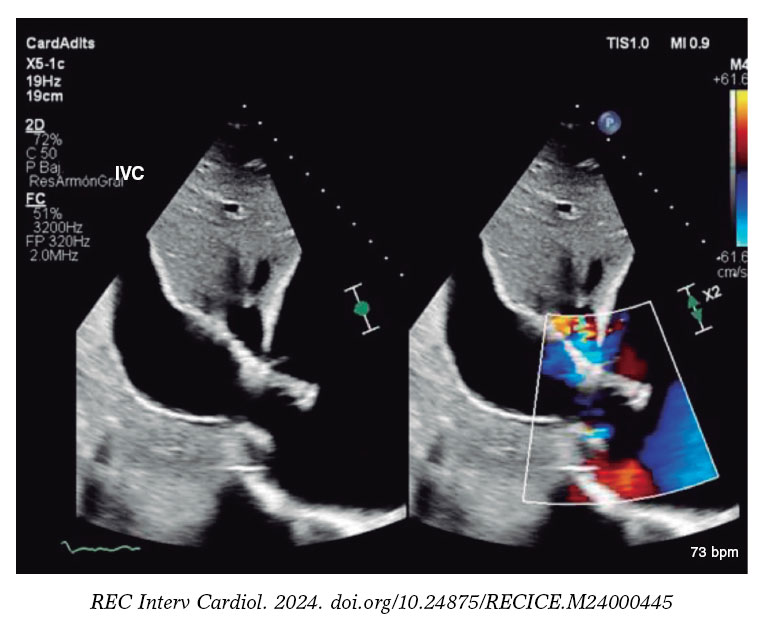
Figure 1.
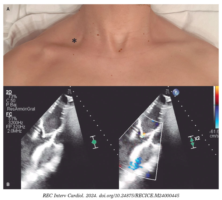
Figure 2.
FUNDING
None declared.
ETHICAL CONSIDERATIONS
Informed consent was obtained from the patient to publish the case.
DECLARATION ON THE USE OF ARTIFICIAL INTELLIGENCE
No artificial intelligence has been used in the preparation of this article.
AUTHORS’ CONTRIBUTIONS
All authors contributed equally to data collection, writing, review, and approval of the article.
CONFLICTS OF INTEREST
A. Pérez de Prado is an associate editor of REC: Interventional Cardiology; the journal’s editorial procedure to ensure impartial processing of the manuscript has been followed. The remaining authors declared no conflicts of interest.
SUPPLEMENTARY DATA
Vídeo 1. Garrote-Coloma C. DOI: 10.24875/RECICE.M24000445
Vídeo 2. Garrote-Coloma C. DOI: 10.24875/RECICE.M24000445


