
This is the case of a 78-year-old man with hypertension and dyslipidemia admitted due to unstable angina. The coronary angiography confirmed the presence of a chronic occlusion at middle left anterior descending coronary artery level and significant stenosis of the middle right coronary artery with a shepherd’s crook morphology of the proximal segment (figure 1A). Angioplasty was performed using an AL-1 catheter and advancing a SION Blue guidewire (Asahi Intecc, Japan). A 4.5 mm × 30 mm zotarolimus-eluting stent was directly implanted. The follow-up angiography revealed the presence of an image consistent with dissection of the artery proximal segment (figure 1B). Afterwards, an intracoronary image was acquired using optical coherence tomography (OCT) (DragonFly OPTIS, Abbott Vascular, United States) that confirmed the presence of a iatrogenic type B dissection presumably due to catheter impaction against the vessel wall. It was treated by implanting a 5.0 mm × 12 mm zotarolimus-eluting stent that overlapped with the previous one. The new OCT performed confirmed its proper expansion and the sealing of dissection. However, a double circle image was seen in several frames (figure 2A,B) gradually coming together until they eventually meet each other (figure 2C). Thanks to the 3D reconstruction of the image, it was revealed that the catheter was folded over itself (figure 3, arrow). The likely mechanism to obtain this image is the difficulty found when trying to advance the OCT catheter due to the double curve created by the withdrawal of the AL-1 catheter (to try to assess the angioplasty result) added to the shepherd’s crook morphology of the artery (zigzag course).
The patient gave his written informed consent for publication purposes.
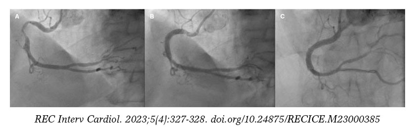
Figura 1
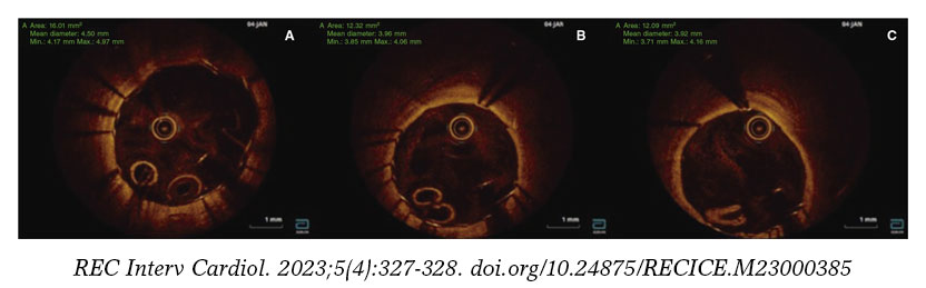
Figura 2
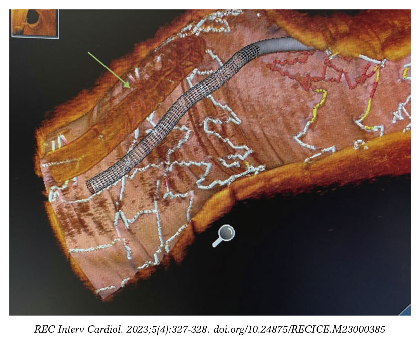
Figura 3
FUNDING
None whatsoever.
AUTHORS’ CONTRIBUTIONS
S. Santos-Martínez, and P. Tejedor-Viñuela drafted the manuscript and completed its critical review. M. Leiva-Gordillo performed the final processing of the images. R. García-Belenger, and P. Morillas-Blasco reviewed the manuscript and approved its final version for publication. All the authors approved such version.
CONFLICTS OF INTEREST
None reported.
* Corresponding author.
E-mail address: sandrasantosmartinez@gmail.com (S. Santos-Martínez).


