Impact Factor: 1.4

Pre-stenting of the right ventricular outflow tract (RVOT) is often performed before percutaneous pulmonary valve implantation to guarantee a stable landing zone. One common off-label indication is the presence of a large native RVOT, which requires the anchoring of an extra-large stent.

This is the case of a 17-year-old woman with a repaired Fallot´s tetralogy admitted for the assessment of severe pulmonary regurgitation with RV dilatation (video 1 of the supplementary data). The patient was ranked as NYHA functional class III. The cardiac magnetic resonance imaging revealed the presence of a large RVOT with diameters of 27 mm x 26 mm (video 2 of the supplementary data).

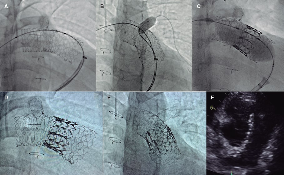
For SAPIEN XT valve implantation (Edwards Lifesciences, United States) and to prevent stent migration, the “double-stent” technique was used. A 57 mm AndraStent XXL stent (Andramed, Germany) was pre-mounted on a 26 mm balloon followed by distal-to-proximal implantation from right pulmonary artery to pulmonary trunk. Afterwards, a second 48 mm AndraStent XXL stent was pre-mounted on a 30 mm balloon and partially implanted overlapping the former stent (figure 1A). A stent that remained in the left pulmonary branch that was implanted at a younger age was dilated with a 10 mm balloon through the new stents (figure 1B). Then, a 29 mm SAPIEN XT valve with 2 extra mL for balloon inflation was advanced and implanted successfully (figure 1C,F). No residual gradient or significant regurgitation were reported. The patient was discharged 2 days later and remains in NYHA class I after 8 months. The father’s patient has given his verbal consent for the publication of this case report.
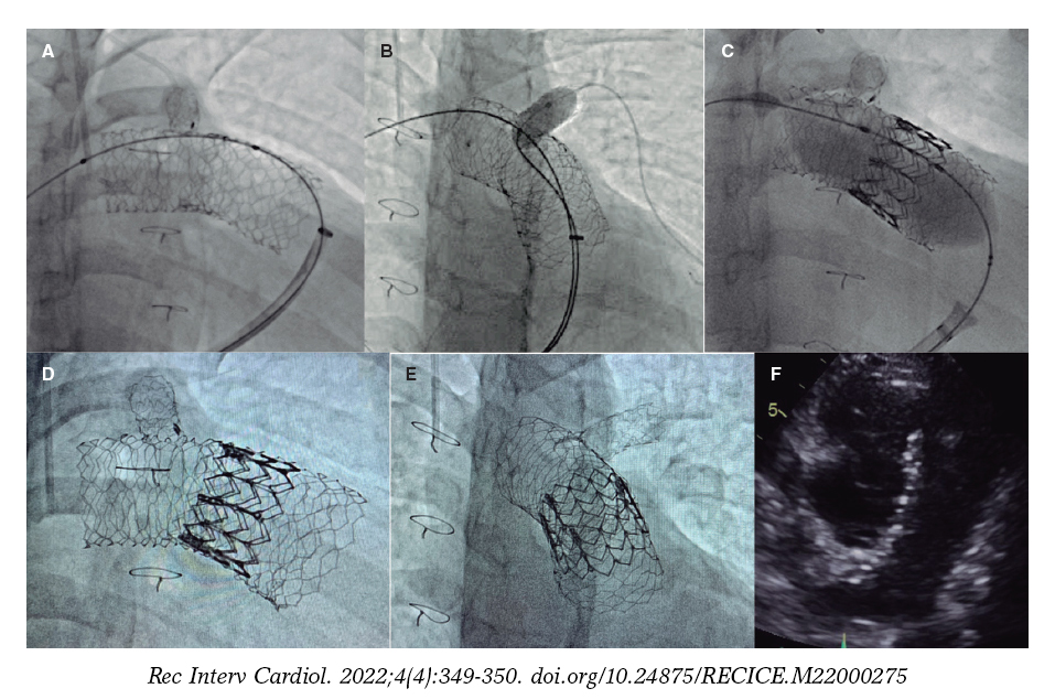

Figure 1.


Double overlapping pre-stenting with the AndraStent XXL stent provides valid anchoring support for percutaneous pulmonary valve implantation in large native RVOTs.

FUNDING

None reported.

AUTHORS’ CONTRIBUTIONS

All the authors drafted this manuscript, read and approved its final version.

CONFLICTS OF INTEREST

None whatsoever.

SUPPLEMENTARY DATA


Vídeo 1. Hernández F. DOI: 10.24875/5/RECICE.M22000275


Vídeo 2. Hernández F. DOI: 10.24875/5/RECICE.M22000275

* Corresponding author.

E-mail address: felipeivus@hotmail.com (F. Hernández Hernández).

The association of Kawasaki disease with the formation of aneurysms and coronary stenoses is well established, especially in gammaglobulinnaïve patients. This is the case of a female patient monitored due to patent ductus arteriosus. The control echocardiography performed at the age of 2 years revealed the presence of giant coronary aneurysms in both coronary arteries. The coronary computed tomography angiography (CCTA) and following cardiac catheterization performed confirmed this finding. The patient’s past medical history revealed she had been admitted at the age of 9 months due to fever compatible with pyelonephritis with good response to antibiotic therapy that could have been consistent with incomplete Kawasaki disease due to fever and further desquamation. Antiplatelet and anticoagulant therapies were administered that, to this date, have been maintained indefinitely.

When the patient was 6 years old, another echocardiography (figure 1: short axis, Ao, aorta, 1A: right arrow: left coronary aneurysm; left arrow: right coronary aneurysm; arrow 1B: right coronary aneurysm), CCTA (figure 2, right arrows: left coronary aneurysm; left arrows: right coronary aneurysm), and cardiac catheterization were performed that revealed the presence of 2 17 mm x 8.8 mm and 7.3 mm × 5.3 mm calcified aneurysms in the right coronary artery (figure 3A,B and videos 1 and 2 of the supplementary data) plus another 8.3 mm × 6.7 mm aneurysm in the left anterior descending coronary artery (figure 3C,D and videos 3 and 4 of the supplementary data) with mild stenosis in the posterior descending artery (figure 3A,B, asterisk). The clinical course revealed no significant size changes compared to diagnosis or complications.


Figure 1.


Figure 2.


Figure 3.


Since the patient had a high cardiovascular risk, after the last cardiac catheterization, treatment with statins was added according to the clinical practice guidelines on the management of Kawasaki disease published by the American Heart Association

To this date, the patient remains asymptomatic, and no complications associated with her disease or treatment have been reported.

The patient’s mother gave her written informed consent to be able to publish this case.

FUNDING

None whatsoever.

AUTHORS’ CONTRIBUTIONS

All authors participated in the drafting of this manuscript, read, and approved its final version.

CONFLICTS OF INTEREST

None reported.

SUPPLEMENTARY DATA


Vídeo 1. Marimón Blanch C. DOI: 10.24875/RECICE.M22000272


Vídeo 2. Marimón Blanch C. DOI: 10.24875/RECICE.M22000272


Vídeo 3. Marimón Blanch C. DOI: 10.24875/RECICE.M22000272


Vídeo 4. Marimón Blanch C. DOI: 10.24875/RECICE.M22000272

* Corresponding author:

E-mail address: cristina.marimon@salutsantjoan.cat (C. Marimón Blanch).

We present the case of a 91-year-old man with a past medical history of moderate-to-severe aortic stenosis referred due to breathlessness. The routine invasive coronary angiography performed before transcatheter aortic valve replacement confirmed the presence of a severely calcified left anterior descending coronary artery proximal segment (figure 1A; video 1 of the supplementary data). The functional assessment performed showed a fractional flow reserve of 0.79. The optical coherence tomography (OCT) interrogation revealed an arc of calcium > 180°, thickness of 0.6 mm, and length > 5 mm with a calcified nodule with a minimum lumen area of 3.49 mm2 (figure 1C; video 2 of the supplementary data). Orbital atherectomy with the Diamondback 360 Coronary OAS (Cardiovascular Systems, Inc., United States) was selected to prepare the lesion. After 5 runs at low speed (80 000 rpm), a new OCT was performed that confirmed the fracture of the calcified nodule achieving a minimum lumen area of 7.5 mm2 (figure 1D; video 3 of the supplementary data). Consecutively, a 2.5 mm x 15 mm SC balloon (OrbusNeich, China) plus a 3.0 mm x 10 mm NC balloon (Medtronic, United States) were advanced through the lesion and inflated twice. Finally, a Megatron 3.5 mm x 20 mm drug-eluting stent (Boston Scientific, United States) was implanted at 20 atm. Both the post-stenting final coronary angiogram (figure 1B; video 4 of the supplementary data) and the OCT pullbacks confirmed the homogeneous expansion and correct apposition of the stent (figure 1E; video 5 of the supplementary data). All the pertinent informed consents were obtained.

Figure 1.

We presented one of the first cases of orbital atherectomy as an effective tool to treat calcified nodules, and the first case of revascularization supported not only by angiography, but also by intracoronary imaging.

FUNDING

None reported.

AUTHORS’ CONTRIBUTIONS

All authors participated in the process of drafting this manuscript. Also, all the authors approved its final version.

CONFLICTS OF INTEREST

None of the authors declared any conflicts of interest in association with this manuscript.

SUPPLEMENTARY DATA


Vídeo 1. Abellas-Sequeiros M. DOI: 10.24875/RECICE.M21000256


Vídeo 2. Abellas-Sequeiros M. DOI: 10.24875/RECICE.M21000256


Vídeo 3. Abellas-Sequeiros M. DOI: 10.24875/RECICE.M21000256


Vídeo 4. Abellas-Sequeiros M. DOI: 10.24875/RECICE.M21000256


Vídeo 5. Abellas-Sequeiros M. DOI: 10.24875/RECICE.M21000256


* Corresponding author: Departamento de Cardiología, Hospital Ramon y Cajal, Carretera Colmenar Viejo Km. 9,100, 28034 Madrid, Spain.

E-mail address: maine.iaca@gmail.com (M. Abellas-Sequeiros).

While performing aortic valve-in-valve procedures the fracture of certain surgical biological valve annuli has been widely reported regarding the implantation of a larger percutaneous valve with a better hemodynamic profile. The pulmonary valve-in-valve technique has not been around that much, but its role is more important because it allows «small» valve implantation in pediatric patients who later progress into significant somatic growths. We present 2 cases after obtaining the express consent of the patients or, if minors, of their legal tutorss.

Case #1 is a 15-year-old patient with Fallot’s tetralogy, and previous surgical implantation of a 19 mm Carpentier Magna Ease valve (Edwards Lifesciences Corp., United States) (internal lumen of 17 mm) at 11 years old. Severe stenosis and moderate regurgitation. The annulus ruptured with a 20 mm x 20 mm Atlas Gold balloon (Becton, Dickinson and Company Franklin Lakes, United States) inflated at 22 atm, and a 23 mm Edwards S3 valve was implanted (figure 1A, angiography of pulmonary artery; figure 1B, rupture with balloon; figure 1C, previous ruptured valve [arrow]; figure 1D, new valve; and videos 1-4 of the supplementary data).


Figure 1.


Case #2 is a 24-year-old patient with Fallot’s tetralogy and previous surgical implantation of a 21 mm Carpentier Edwards valve (internal lumen of 19 mm) at 14 years old. Moderate doble lesion. The valve ruptured with a 24 mm x 20 mm Atlas Gold balloon inflated at 20 atm, and a 26 mm Edwards XT valve was implanted (figure 2A, angiography of pulmonary artery; figure 2B, rupture with balloon; figure 2C, previous ruptured valved distended with a 25 mm Cristal balloon (Balt, France) [arrow]; figure 2D, new valve; and videos 5-8 of the supplementary data).


Figure 2.


The rupture of a biological prosthetic valve annulus in pulmonary position facilitates the implantation of another larger valve to match the growth of each patient, and makes valve-in-valve procedures possible in the future. This procedure can be unnecessary if the first prosthetic valve implantation is performed with long-term thinking and the largest possible valve is used.

FUNDING

None whatsoever.

AUTHORS’ CONTRIBUTIONS

The 3 authors participated in the idea, writing, and review of this article.

CONFLICTS OF INTEREST

None reported.

SUPPLEMENTARY DATA


Vídeo 1. Betrián Blasco P. DOI: 10.24875/RECICE.M21000253


Vídeo 2. Betrián Blasco P. DOI: 10.24875/RECICE.M21000253


Vídeo 3. Betrián Blasco P. DOI: 10.24875/RECICE.M21000253


Vídeo 4. Betrián Blasco P. DOI: 10.24875/RECICE.M21000253


Vídeo 5. Betrián Blasco P. DOI: 10.24875/RECICE.M21000253


Vídeo 6. Betrián Blasco P. DOI: 10.24875/RECICE.M21000253


Vídeo 7. Betrián Blasco P. DOI: 10.24875/RECICE.M21000253


Vídeo 8. Betrián Blasco P. DOI: 10.24875/RECICE.M21000253

* Corresponding author: Paseo de Valle de Hebrón 119-129, 08035 Barcelona, Spain.

E-mail address: pedrobetrian@yahoo.es (P. Betrián Blasco).

This is the case of a 34-year-old woman treated percutaneously for aortic coarctation repair. The patient’s past medical history included muscular ventricular septal defects with spontaneous closure, bicuspid aortic valve with asymptomatic moderate regurgitation with normal ventricular volume and function, and juxtaductal coarctation of the aorta treated with patch aortoplasty at 2 months old.

The data of progressive recoarctation were obtained from the echocardiography and magnetic resonance imaging at the outpatient follow-up, and the hypertensive response from the ergometric work. The case was brought to the heart team that decided to perform endovascular treatment. The aortography performed via right femoral access confirmed the presence of recoarctation at the junction between the aortic arch and the descending thoracic aorta (figure 1A,B, blue arrow) with a minimum diameter of 9 mm, and a peak-to-peak gradient of 20 mmHg. Similarly, a saccular-shaped pseudoaneurysm was found adjacent to the stenosed region (figure 1A,B; figure 2A, yellow arrow; video 1 of the supplementary data) with internal diameters of 8 mm x 10 mm (figure 2B). Therefore, it was decided to implant a 45 mm long covered stent (Covered CP Stent, NuMED Canada) (figure 3) with good angiographic results (videos 2 and 3 of the supplementary data), no evidence of residual gradient, and successful and complete exclusion of the pseudoaneurysm.


Figure 1.


Figure 2.


Figure 3.


The use of imaging modalities with 3D analysis including traditional fluoroscopy is a very useful tool to diagnosis of complex anatomies and plan therapeutic alternatives. Also, it allows us to shorten the duration of the procedure, reduce the dose of contrast used, and minimize radiation exposure time.

The patient’s written informed consent was obtained before publishing this clinical case.

FUNDING

None whatsoever.

AUTHORS’ CONTRIBUTIONS

N. Barja González wrote the manuscript first draft as well as its final version. F. Rueda Núñez, and I. Martínez Bendayán provided the supplementary data and reviewed the manuscript draft and final version.

CONFLICTS OF INTEREST

None reported.

SUPPLEMENTARY DATA


Vídeo 1. Barja González N. DOI: 10.24875/RECICE.M21000247


Vídeo 2. Barja González N. DOI: 10.24875/RECICE.M21000247


Vídeo 3. Barja González N. DOI: 10.24875/RECICE.M21000247


* Corresponding author: Av. Roma s/n, 33011 Oviedo, Asturias, Spain.

E-mail address: noeminbg@gmail.com (N. Barja González).

Editorials

Recic Uk 23 087 F2

Original articles

Recic 23 026 Uk F1

Editorials


Original articles

Recic 23 007 Uk F1
Recic Uk 23 053 F3
Recic Uk 23 042 F3

Debate

Debate: Asymptomatic severe aortic stenosis: when should we intervene?

Recic 23 082 F1
Recic 23 083 F1