ABSTRACT
Introduction and objectives: Infective endocarditis (IE) is a rare but serious complication in patients with aortic valve stenosis undergoing transcatheter aortic valve implantation (TAVI). The spread of this technique to lower risk patients means that this complication may increase. The objective of this study was to analyze the incidence and mortality of IE in TAVI patients vs patients undergoing surgical aortic valve replacement (SAVR).
Methods: We conducted an observational, single-center, retrospective cohort study that included all cases of IE diagnosed consecutively in a Spanish reference center from 2008 through 2022 in patients with TAVI vs SAVR.
Results: The study included a total of 10 cases of IE in 778 patients treated with TAVI, with an incidence rate of 0.09/100 patients/year vs an incidence rate of 0.12/100 patients/year in surgical bioprostheses with 24 cases in 1457 patients (P = .64) (median follow-up of 49 months (p25-p75: 29-108). Clinical features were very similar, with 50% of TAVI patients having cardiac complications vs 33% of SAVR patients (P = .33). Although 40% of the patients from the TAVI group had a surgical indication for IE and 50% for SAVR, P = .49), only half of them underwent surgery in both groups (20% TAVI vs 25% SAVR; P = .93). No differences were reported in the 1-year mortality rate (30% TAVI vs 29% SAVR; P = .56).
Conclusions: The incidence rate of IE in this long series of TAVI patients was low and despite the worse clinical profile of TAVI patients, no significant mortality differences were found compared with the group of patients with surgical bioprosthesis.
Keywords: Infectious endocarditis. Aortic stenosis. Surgical aortic valve replacement. Transcatheter aortic valve implantation.
RESUMEN
Introducción y objetivos: La endocarditis infecciosa (EI) es una complicación infrecuente, pero grave, en los pacientes con estenosis valvular aórtica que han recibido un implante percutáneo de válvula aórtica (TAVI). La extensión de esta técnica a pacientes de menor riesgo hace que esta complicación pueda aumentar. El objetivo del estudio fue analizar la incidencia y la mortalidad de la EI en pacientes con TAVI en comparación con la EI en pacientes con recambio valvular aórtico (RVAo).
Métodos: Estudio observacional, unicéntrico, retrospectivo de cohortes, que incluyó todos los casos de EI diagnosticados de manera consecutiva en un centro español de referencia, desde 2008 hasta 2022, en pacientes con TAVI, y se compararon con las EI en pacientes con RVAo.
Resultados: Hubo 10 casos de EI en 778 pacientes tratados con TAVI, con una tasa de incidencia de 0,09/100 pacientes/año, frente a 24 casos en 1.457 pacientes con RVAo, con una tasa de incidencia de 0,12/100 pacientes/año (p = 0,64), en una mediana de seguimiento de 49 meses (p25-p75: 29-108). Los pacientes con TAVI eran mayores, tenían más diabetes mellitus y un EuroSCORE mayor. El microorganismo más frecuente fue el enterococo (30% TAVI frente a 33% RVAo; p = 0,89). La evolución clínica fue muy similar, con un 50% de pacientes con TAVI que tuvieron una complicación cardiaca frente al 33% de los pacientes con RVAo (p = 0,33). En el grupo de TAVI, el 40% tuvieron indicación quirúrgica por la EI, frente al 50% en el grupo de RVAo (p = 0,49), pero solo la mitad fueron intervenidos en ambos grupos (20% TAVI frente a 25% RVAo; p = 0,93). No hubo diferencias en la mortalidad al año (30% TAVI frente a 29% RVAo; p = 0,56).
Conclusiones: La incidencia de EI en esta serie de pacientes con TAVI fue baja, y pese a un peor perfil clínico en los pacientes con TAVI, no se encontraron diferencias significativas en la mortalidad con el grupo de pacientes con RVAo.
Palabras clave: Endocarditis infecciosa. Estenosis aórtica. Recambio valvular aórtico. Implante percutáneo de válvula aórtica.
Abbreviations
IE: infectious endocarditis. SAVR: surgical aortic valve replacement. TAVI: transcatheter aortic valve implantation.
INTRODUCTION
Transcatheter aortic valve implantation (TAVI) revolutionized the treatment of severe aortic valve stenosis over the past decade.1,2 Furthermore, in recent years, there has been a clear preference for TAVI over surgical techniques regarding the treatment of this valvular heart disease, with an increasing number of patients, including low surgical risk ones.3 Although post-TAVI infective endocarditis (IE) is a rare complication reported in 1% up to 6% of patients, it is often associated with grim clinical outcomes and high mortality rates despite diagnostic1,2 and therapeutic4 advances. Complications are expected to rise exponentially as the number of TAVIs continues to rise at a constant rate in our setting as well.5
Few studies have compared the incidence of IE on TAVI and surgical aortic valve replacement (SAVR). However, randomized clinical trials have shown similar annual incidences of IE after SAVR and TAVI.6,7 Due to the strict patient selection of such studies, the results obtained in each center during the routine clinical practice may vary, and few studies like this using dedicated databases have been conducted. A multicenter study8 revealed the characteristics of IE on TAVI vs SAVR and the outcomes of a large patient cohort, yet it did not analyze the incidence of IE. Assessing the outcomes of this severe complication at each center should be a priority due to its potential impact on decision-making for the heart team.3
The aim of the present study was to analyze both the incidence and mortality rates of IE on TAVI vs SAVR patients from a tertiary teaching hospital in a long series of patients with severe aortic stenosis.
METHODS
Study design and population
We conducted an observational, single-center, retrospective study of a prospective cohort including all cases of IE in symptomatic patients with severe aortic valve stenosis treated with TAVI or SAVR using a biological prosthetic valve, diagnosed, and followed at a tertiary teaching referral hospital by an endocarditis team from January 2008 through December 2022.
Endpoints and definitions
The primary endpoint of the study was to analyze the overall incidence and mortality rates of IE on TAVI at our center. Secondary endpoint was to compare both rates in patients with IE on TAVI and SAVR during the same period. Another secondary endpoint was to study the number of patients who underwent surgery for IE in both groups. IE was diagnosed using the Duke criteria9 or the 2015 European Society of Cardiology modified criteria10 depending on the time of diagnosis in the study period. Cases of IE in TAVI patients during this period were identified, and their characteristics were compared with those of IE in SAVR patients.
Follow-up
Follow-up events were defined based on the criteria established by the Valve Academic Research Consortium-2.11 All complications, need for surgery, and mortality at the follow-up were recorded. IE-related cardiac complications included decompensated heart failure, fistula, prosthetic dehiscence, abscess, and complete atrioventricular block. Systemic complications included acute kidney failure, sepsis, embolism, and disorders of the central nervous system. Early IE was defined as that occurring within the first year after TAVI or surgery based on European recommendations.10 Since 1987, a prospective protocol for inclusion and follow-up of all IE cases has been in place at our center, with a systematic registry including in-person visits, at least, annually for all patients, as well as phone consultations when needed.
Statistical analysis
Qualitative data are expressed as percentages, and continuous data as mean and standard deviation or median [interquartile range], depending on whether they follow a normal distribution. Inter-group comparisons were drawn using the chi-square test or Fisher’s exact test for qualitative variables, and the Student’s t-test or Mann-Whitney U test for continuous variables, as appropriate. Time-to-event analyses for all-cause mortality were conducted using Kaplan-Meier curves. All tests were two-sided, and P values < .05 were considered statistically significant. Statistical analyses were performed with SPSS software (version 24; IBM Corp, Armonk, NY, United States).
RESULTS
Study population and incidence rate
During the study period, a total of 778 patients successfully underwent TAVI, 70% of them with self-expanding valves. After a median follow-up of 49 months (p25-p75, 29–108 months), 10 cases of IE were eventually diagnosed, which amounts to an overall incidence rate of 1.29% (an incidence rate of 0.09/100 patient-years). Twenty-four of the 1457 patients treated with surgical bioprostheses were diagnosed with IE for an overall incidence rate of 1.64% (an incidence rate of 0.12/100 patient-years). The hazard ratio for the IE incidence rate between the 2 groups was 0.75 (95% confidence interval, 0.36–1.57). The incidence rate of IE on TAVI remained stable throughout the study period, as did the incidence of IE on surgical bioprostheses. Four of the 10 IE cases in TAVI occurred between 2008 and 2015—incidence of 1.21%—and 6 between 2016 and 2022 (incidence of 1.33%). The incidence rates of IE on surgical bioprostheses for the same periods were 1.62% and 1.67%, respectively. Clinical characteristics, treatment, and mortality rates were also similar across periods in both IE groups.
Characteristics of IE in TAVI and SAVR
Approximately half of the cases reported in both groups turned out to be early IE: 5 of the 10 TAVI IE cases and 11 of the 24 cases described on surgical bioprostheses occurred within the first year after implantation (50% TAVI and 46% SAVR; P = .56). The remaining cases were late prosthetic IE (table 1). Among TAVI IE cases, the 5 early ones were diagnosed 2, 4, 6, 8, and 11 months after implantation, while the 5 late ones were diagnosed within years 2 (3 cases) and 3 (2 cases). Among the IE reported on surgical bioprostheses, early cases were diagnosed within the first 2 months (2 cases), between months 3 and 6 (3 cases), and between months 6 and 12 (5 cases) after surgery. Regarding late cases, 5 occurred in year 2, 4 in year 3, and 4 > 3 years after surgery.
Table 1. Baseline characteristics of patients with infectious endocarditis after transcatheter aortic valve implantation or surgical aortic valve replacement
| Baseline characteristics | Total (n = 34) | TAVI (n = 10) | SAVR (n = 24) | P |
|---|---|---|---|---|
| Age (years) | 67 (53-81) | 76 (67-85) | 63 (49-77) | .001 |
| Female | 13 (38%) | 4 (40%) | 9 (37%) | .594 |
| Hypertension | 28 (82%) | 10 (100%) | 18 (75%) | .100 |
| Type 2 diabetes mellitus | 14 (41%) | 7 (70%) | 7 (29%) | .034 |
| COPD | 8 (33%) | 3 (30%) | 5 (21%) | .435 |
| CKD (GFR < 60) | 12 (35%) | 3 (30%) | 9 (38%) | .498 |
| Atrial fibrillation | 17 (50%) | 7 (70%) | 10 (42%) | .129 |
| Ischemic heart disease | 4 (12%) | 1 (10%) | 3 (12%) | .666 |
| Functional class II/III | 18 (53%) | 6 (60%) | 12 (50%) | .488 |
| EuroSCORE II | 7.41 ± 4.1 | 3.6 ± 2.8 | .007 | |
|
CKD, chronic kidney disease; COPD, chronic obstructive pulmonary disease; GFR, glomerular filtration rate; SAVR, surgical aortic valve replacement; TAVI, transcatheter aortic valve implantation. |
||||
A possible source of infection different from implantation or surgery was identified in 2 of the 10 TAVI IE cases (20%) and in 2 of the 24 surgical bioprostheses. IE cases (8.3%). Only 1 of the early IE cases was associated with a possible infection source: a colonoscopy in a TAVI IE patient due to Enterococcus. None of the SAVR IE cases had an identified source, with implantation or the perioperative period being regarded as the probable sources of infection in the remaining patients. Among late IE cases, 1 TAVI IE case was associated with a dental procedure—despite proper antibiotic prophylaxis—and 2 SAVR IE cases with an upper gastrointestinal endoscopy and a dental visit, respectively. These patients’ clinical characteristics are shown in table 1. TAVI patients were older, with a median [interquartile range] age of 76 years [67–85] vs 63 years [49–77] of those treated with surgical bioprostheses (P < .001) and had higher rates of diabetes (70% vs 29%; P = .034). However, there were no significant differences in sex, other comorbidities, or symptoms. As expected, the TAVI group had a significantly higher EuroSCORE II vs the SAVR group (table 1). The profile of causative pathogens was very similar between the 2 groups (table 2). Enterococci were the pathogens most widely identified, followed by coagulase-negative staphylococci and Staphylococcus aureus, also with no significant differences being reported between the 2 groups. In 17% of SAVR patients and 20% of TAVI patients, the causative agent could not be identified (figure 1). Diagnostic echocardiographic findings of IE were also very similar between the 2 groups: transthoracic echocardiography identified IE only in half of the TAVI cases vs 37% of the cases treated with surgical bioprostheses (
Table 2. Microbiological profile of the most common microorganisms and diagnostic lesions in the echocardiogram of patients with infectious endocarditis after transcatheter aortic valve implantation or surgical aortic valve replacement
| Microbiological profile and echocardiogram | Total (n = 34) | TAVI (n = 10) | SAVR (n = 24) | P |
|---|---|---|---|---|
| Early IE (< 1 year) | 16 (47%) | 5 (50%) | 11 (46%) | .560 |
| Microorganism | ||||
| Enterococcus | 11 (32%) | 3 (30%) | 8 (33%) | .891 |
| Staphylococcus epidermidis | 9 (26%) | 3 (30%) | 6 (25%) | .819 |
| Staphylococcus aureus | 4 (12%) | 1 (10%) | 3 (12%) | .854 |
| Other/Unknown | 10 (29%) | 3 (30%) | 7 (29%) | .153 |
| Lesion in echocardiogram | ||||
| TTE | 14 (41%) | 5 (50%) | 9 (37%) | .382 |
| TEE | 26 (76%) | 10 (100%) | 16 (67%) | .101 |
|
IE, infectious endocarditis; TEE, transesophageal echocardiogram; TTE, transthoracic echocardiogram; SAVR, surgical aortic valve replacement; TAVI, transcatheter aortic valve implantation. |
||||
Disease progression
The course of the disease was similar in the 2 groups (table 3). Half of the TAVI group experienced cardiac complications, as did one-third of the SAVR group, without any significant differences being reported. Half of the patients had a surgical indication due to IE (40% in the TAVI group vs 50% in the SAVR group; P = .49), but among those, only 20% of the TAVI group and 25% of the SAVR group eventually underwent surgery (P = .93) (table 3). The in- hospital mortality rate was similar (20% in the TAVI group vs 25% in the SAVR group; P = .51), without any significant differences being reported in the 1-year mortality rate, which remained high—at approximately 30%—in both groups (table 3, figure 1, and figure 3).
Table 3. Complications, rate of surgical procedures, and 1-year mortality rate in patients with infectious endocarditis after transcatheter aortic valve implantation or surgical aortic valve replacement
| Complications and mortality | Total (n = 34) | TAVI (n = 10) | SAVR (n = 24) | P |
|---|---|---|---|---|
| Cardiac complications | 13 (38%) | 5 (50%) | 8 (33%) | .329 |
| Systemic complications | 15 (44%) | 2 (20%) | 13 (54%) | .072 |
| Indication for surgery | 16 (47%) | 4 (40%) | 12 (50%) | .491 |
| Surgery performed | 8 (23%) | 2 (20%) | 6 (25%) | .932 |
| Surgery not performed | 26 (77%) | 8 (80%) | 18 (75%) | .909 |
| 1-year mortality | 10 (29%) | 3 (30%) | 7 (29%) | .562 |
| In-hospital mortality | 8 (24%) | 2 (20%) | 6 (25%) | .512 |
|
SAVR, surgical aortic valve replacement; TAVI, transcatheter aortic valve implantation. |
||||

Figure 1. Infective endocarditis (IE) after transcatheter aortic valve implantation in a cohort of 778 patients vs a cohort of patients with surgical bioprostheses-related IE. SAVR, surgical aortic valve replacement; TAVI, transcatheter aortic valve implantation; TEE, transesophageal echocardiography; TTE, transthoracic echocardiography.

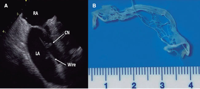
Figure 2. Transesophageal echocardiogram showing a large vegetation on the leaflets of a transcatheter aortic valve (arrows).

Figure 3. Kaplan-Meier curves showing the 1-year mortality rate in patients with infective endocarditis after transcatheter aortic valve implantation (TAVI) or surgical aortic valve replacement (SAVR).
DISCUSSION
The main finding of our study was that the incidence rate of IE on TAVI is low in our setting and similar to that of surgical aortic bioprostheses, despite being patients with higher surgical risk, older age, and more comorbidities. These results are similar to those reported in the literature (1% up to 6%);4 however, more recent large TAVI trials suggest lower rates. The PARTNER 3 trial reported annual rates of 0.2%,1 and similarly, the low-risk Evolut study2 found incidence rates of 0.1% and 0.2% at 30 days and 1 year, respectivelu, which is more consistent with our rates. Studies directly comparing the incidence of IE after SAVR and TAVI are scarce, and some have produced contradictory results.7,12 However, most observational studies based on large national databases and randomized clinical trials have found no statistically significant differences on this regard,6,13,14 even though TAVI patients are older and generally have more comorbidities, which is consistent with our results. There were also no differences in our study regarding early IE incidence (50%), which was similar in both groups, although slightly lower for IE on TAVI vs what has been reported by the literature (rates up to 64%.)15 However, a multicenter study conducted in Spain found a higher rate of early IE in TAVI vs SAVR patients (78.1% vs 39.3%; P = .001), which is significantly higher than that described in our series for the TAVI group.16 This could be explained by the different antibiotic prophylaxis regimens and procedures used at each center.
Secondly, another relevant finding from our study is that enterococci were the main cause of IE in both patient groups, which is consistent with the literature on TAVI-related IE17,18 but not on surgical bioprostheses-related IE, of which S. aureus is usually the main microorganism involved. These differences are difficult to explain, as the increase in enterococcal incidence as a causal agent in TAVI is primarily associated with older patient age and transfemoral access but still would not explain why it is also the most common pathogen in SAVR-related IE.4 Compared with a multicenter Spanish series of similar characteristics to ours,16 there are some differences: the most common causative microorganism of IE—in both TAVI and SAVR— was Staphylococcus epidermidis, followed by enterococci— less frequent than in our series—and thirdly, S. aureus. These differences could be explained by varying antibiotic prophylaxis regimens used in different settings, underscoring the critical importance of understanding the most common microorganisms involved in prosthetic IE, in general, to apply the most appropriate and effective prophylaxis regimen.
Another noteworthy aspect of this study is that echocardiographic lesions diagnostic of IE could be identified in 100% of TAVI- related IE cases vs 67% of SAVR-related IE cases in surgical prostheses. This contrasts sharply with most reports, which indicate that the combined sensitivity rate of transthoracic and transesophageal echocardiography was 67.8% in TAVI patients, 73% in SAVR patients, and nearly 90% in native valves.19 However, a very recent study comparing patients with IE after TAVI or SAVR confirmed that vegetations were identified via echocardiogram in up to 82% of the TAVI group, more in line with our findings and significantly higher than the diagnostic rate of IE reported in surgical prostheses (62.5%; P < .001).8 Our results also differ from those reported in the literature in that most lesions found via echocardiography were vegetations, whereas other authors, such as Salaun et al.,20 reported vegetations in only 5 out of 11 cases diagnosed with TAVI-related IE, with the remaining cases being atypical lesions.
Lastly, this study also highlights the course of the disease, with a 1-year mortality rate of 30%, which is lower than that reported in other published studies (between 33% and 66%).4,12,21-23 The in-hospital mortality rate was also lower than that reported in other studies, such as the international multicenter registry of post-TAVI endocarditis21 (36% vs 20% in our series) and much lower than the Spanish multicenter trial (35%).16 These differences may be due to the wide variability in patient characteristics across studies. Of note, there were no significant differences in mortality when comparing surgical bioprostheses-related IE, unlike other series reporting lower mortality for the latter vs TAVI-related IE.23 However, a study published by Panagides et al.,8 comparing early and 1-year mortality in IE after TAVI and SAVR using propensity score matching found no significant differences between the 2. In our series, only 20% of TAVI patients with a surgical indication underwent surgery, which is consistent with other studies where surgical rates were similar to ours, with optimal medical therapy being the most common strategy.4,12,16,22,23 Some studies found no improvement in prognosis for these patients undergoing surgery, with similar mortality rates associated with optimal medical therapy,24 while a meta-analysis published by Tinica et al.25 showed that the surgical strategy was significantly superior to conservative therapy. In any case, given the lack of strong evidence, the treatment strategy for TAVI-related IE remains unclear, even if the presence of complications warrants surgical indication, and decisions should be based on local expertise.
Study limitations
Our study has the inherent limitations of its observational design, with data collected over many years, during which diagnostic criteria for IE, valve types—towards better technical models—and procedural approaches—towards less invasive methods—have evolved. The small number of IE cases in the 2 groups means results should be interpreted with caution. As this is a crude analysis, the presence of confounding bias cannot be ruled out; nevertheless, data show real-life outcomes. Additionally, patients with mechanical valves were not included, so results cannot be extrapolated to this group.
CONCLUSIONS
In our setting, TAVI-related IE has a low overall incidence rate, with no significant differences vs SAVR-related IE, despite involving older and more comorbid patients. Regarding the causative microorganism of IE—both in surgical and percutaneous surgical bioprostheses—enterococci were the most common pathogen. There were no differences in mortality between the 2 types of aortic valves, and treatment for IE was predominantly conservative.
FUNDING
None declared.
ETHICAL CONSIDERATIONS
The study was conducted in full compliance with the Declaration of Helsinki and approved by the Clinical Research Ethics Committee at the beginning of the registry in 1987. However, for patients with TAVI or SAVR, all participants signed an informed consent form authorizing the collection and analysis of their data for research purposes. Since this was an observational study, patient treatment was not impacted. For this new registry analysis, approval was obtained from our center ethics committee. Data were handled completely anonymously in full compliance with the provisions of Organic Law 3/2018 of the Spanish Data Protection Authority.
STATEMENT ON THE USE OF ARTIFICIAL INTELLIGENCE
No artificial intelligence was used in the development of this work.
AUTHORS’ CONTRIBUTIONS
A. Roldán participated in data collection and analysis. C. Urbano participated in data collection and analysis. N. Aguayo participated in patient identification, data collection, and analysis. M. Crespín, J. López, and J.C. Castillo participated in the conception of the article and its interpretation. R. González contributed to statistical analysis and result interpretation. D. Mesa and M. Ruiz contributed to data processing, analysis and result interpretation, and collaborated in the revision and preparation of the manuscript for publication. J. Perea, I. Gallo, J. Suárez de Lezo, and S. Ojeda participated in the conception of the work, helped gather patient information, and provided guidance on literature review and manuscript drafting. M. Pan and M. Anguita supervised all stages of manuscript drafting, from conception, data collection, and result interpretation, to revision, correction, and preparation of the article for submission.
CONFLICTS OF INTEREST
S. Ojeda is an associate editor of REC: Interventional Cardiology. The journal’s editorial procedure to ensure impartial handling of the manuscript has been followed. The remaining authors declared no conflicts of interest whatsoever.
WHAT IS KNOWN ABOUT THE TOPIC?
- TAVI-related IE is a rare complication associated with high morbidity and mortality rates. Few studies have compared TAVI- and SAVR-related IE; however, despite sometimes contradictory results, published data generally show similar figures for incidence and mortality. Given the expansion of TAVI indications in recent years to younger and lower-risk patients, it is essential to understand outcomes in different settings.
WHAT DOES THIS STUDY ADD?
- Understanding the reality of a reference center in the management of aortic stenosis using different techniques, such as TAVI and SAVR with surgical bioprosthesis—particularly regarding incidence and mortality rates in a large patient series—is essential for selecting the most appropriate treatment. The low incidence of IE in this study, along with mortality rates consistent with or lower than those published in the literature, helps the heart team make appropriate decisions. Finally, identifying the most common pathogens causing IE in our setting is critical for establishing the most effective prophylactic protocols.
REFERENCES
1. Popma JJ, Deeb GM, Yakubov SJ, et al. Evolut Low Risk Trial Investigators. Transcatheter Aortic-Valve Replacement with a Self-Expanding Valve in Low-Risk Patients. N Engl J Med. 2019;380:1706-1715.
2. Mack MJ, Leon MB, Thourani VH, et al. PARTNER 3 Investigators. Transcatheter Aortic-Valve Replacement with a Balloon-Expandable Valve in Low-Risk Patients. N Engl J Med. 2019;380:1695-1705.
3. Vahanian A, Beyersdorf F, Praz F, et al. 2021 ESC/EACTS Guidelines for the management of valvular heart disease:Developed by the Task Force for the management of valvular heart disease of the European Society of Cardiology (ESC) and the European Association for Cardio-Thoracic Surgery (EACTS). Eur Heart J. 2022;43:561-632.
4. Del Val D, Panagides V, Mestres CA, MiróJM, Rodés-Cabau J. Infective Endocarditis After Transcatheter Aortic Valve Replacement:JACC State-of-the-Art Review. J Am Coll Cardiol. 2023;81:394-412.
5. Íñiguez-Romo A, Zueco-Gil JJ, Álvarez-BartoloméM, et al. Outcomes of transcatheter aortic valve implantation in Spain through the Activity Registry of Specialized Health Care. REC Interv Cardiol. 2022;4:123-131.
6. Summers MR, Leon MB, Smith CR, et al. Prosthetic valve endocarditis after TAVR and SAVR. Circulation. 2019;140:1984-1994.
7. Ando T, Ashraf S, Villablanca PA, et al. Meta-analysis comparing the incidence of infective endocarditis following transcatheter aortic valve implantation versus surgical aortic valve replacement. Am J Cardiol. 2019;123:827-832.
8. Panagides V, Cuervo G, Llopis J, et al. TAVI Infective Endocarditis International Registry and ICE Investigators. Infective Endocarditis After Transcatheter Versus Surgical Aortic Valve Replacement. Clin Infect Dis.2024;78:179-187.
9. Li JS, Sexton DJ, Mick N, et al. Proposed modifications to the Duke criteria for the diagnosis of infective endocarditis. Clin Infect Dis. 2000;30:633-638.
10. Habib G, Lancellotti P, Antunes MJ, et al. 2015 ESC Guidelines for the management of infective endocarditis:The Task Force for the Management of Infective Endocarditis of the European Society of Cardiology (ESC). Endorsed by:European Association for Cardio-Thoracic Surgery (EACTS), the European Association of Nuclear Medicine (EANM). Eur Heart J. 2015;36:3075-3128.
11. Kappetein AP, Head SJ, Généreux P, et al. Updated standardized endpoint definitions for transcatheter aortic valve implantation:the Valve Academic Research Consortium-2 consensus document. J Am Coll Cardiol. 2012;60:1438-1454.
12. Cahill TJ, Raby J, Jewell PD, et al. Risk of infective endocarditis after surgical and transcatheter aortic valve replacement. Heart. 2022;108:639-647.
13. Kolte D, Goldsweig A, Kennedy KF, et al. Comparison of incidence, predictors, and outcomes of early infective endocarditis after transcatheter aortic valve implantation versus surgical aortic valve replacement in the United States. Am J Cardiol. 2018;122:2112-2119.
14. Moriyama N, Laakso T, Biancari F, et al. Prosthetic valve endocarditis after transcatheter or surgical aortic valve replacement with a bioprosthesis:results from the FinnValve registry. EuroIntervention. 2019;15:e500-e507.
15. Mentias A, Girotra S, Desai MY, et al. Incidence, predictors, and outcomes of endocarditis after transcatheter aortic valve replacement in the United States. J Am Coll Cardiol Intv. 2020;13:1973-1982.
16. Jerónimo A, Olmos C, Zulet P, et al. Clinical characteristics and outcomes of aortic prosthetic valve endocarditis:comparison between transcatheter and surgical bioprostheses. Infection. 2024. https://doi.org/10.1007/s15010-024-02302-0.
17. Del Val D, Abdel-Wahab M, Linke A, et al. Temporal trends, characteristics, and outcomes of infective endocarditis after transcatheter aortic valve replacement. Clin Infect Dis. 2021;73:e3750-e3758.
18. Strange JE, Østergaard L, Køber L, et al. Patient Characteristics, Microbiology, and Mortality of Infective Endocarditis After Transcatheter Aortic Valve Implantation. Clin Infect Dis. 2023;77:1617-1625.
19. Wang A, Athan E, Pappas PA, et al. International Collaboration on Endocarditis–Prospective Cohort Study Investigators. Contemporary clinical profile and outcome of prosthetic valve endocarditis. JAMA. 2007;297:1354-1361.
20. Salaun E, Sportouch L, Barral P-A, et al. Diagnosis of infective endocarditis after TAVR:value of a multimodality imaging approach. JACC Cardiovasc Imaging. 2018;11:143-146.
21. Del Val D, Linke A, Abdel-Wahab M, et al. Long-term outcomes after infective endocarditis after transcatheter aortic valve replacement. Circulation. 2020;142:1497-1499.
22. Amat-Santos IJ, Messika-Zeitoun D, Eltchaninoff H, et al. Infective endocarditis after transcatheter aortic valve implantation:Results from a large multicenter registry. Circulation. 2015;131:1566-1574.
23. Regueiro A, Linke A, Latib A, et al. Association between transcatheter aortic valve replacement and subsequent infective endocarditis and in-hospital death. JAMA. 2016;316:1083-1092.
24. Mangner N, Del Val D, Abdel-Wahab M, et al. Surgical treatment of patients with infective endocarditis after transcatheter aortic valve implantation. J Am Coll Cardiol. 2022;79:772-785.
25. Tinica G, Tarus A, Enache M, et al. Infective endocarditis after TAVI:a meta-analysis and systematic review of epidemiology, risk factors and clinical consequences. Rev Cardiovasc Med. 2020;21:263-274.


