
Innovation in interventional cardiology
High-definition intravascular ultrasound
30/04/2019
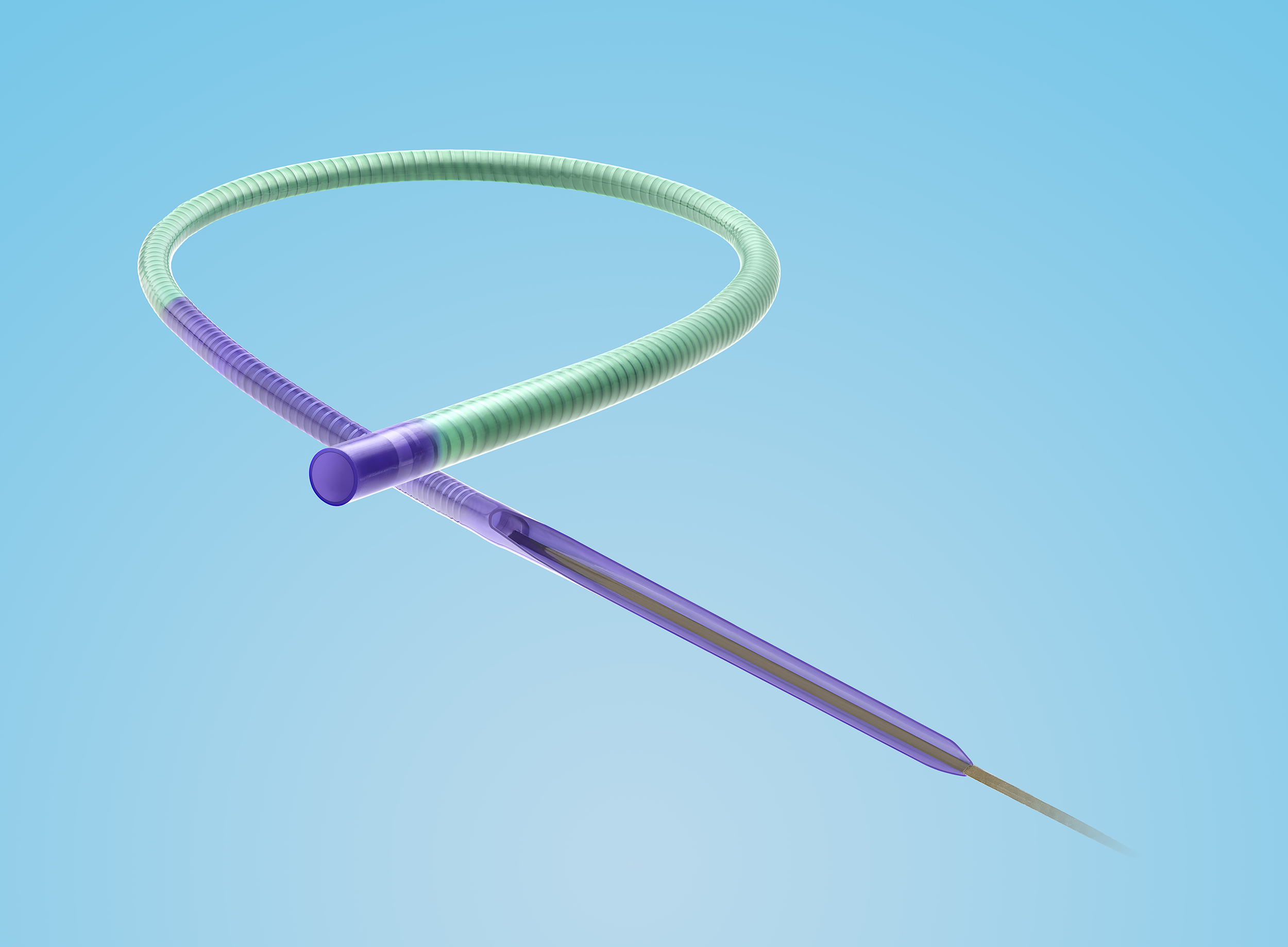
A new guide extension catheter named Telescope (Medtronic) has just been released on the market, and we have already tested it. Its advantage over other available extension catheters is that its structure differs to allow a highly safe and effective catheter release in distal portions of the coronary arteries.
The characteristics of the different parts of the device are as follows:
- Distal segment. The catheter has a coil-based structure, unlike others which contain mesh. The distal portion is more flexible than the proximal portion. The polymer tip measures 2 mm (it has a radiopaque marker) and is produced by extrusion; its flexibility allows deflections that adapt appropriately to the tortuosity of the coronary artery, to calcifications, and to lesions encountered in the coronary lumen, without causing dissection.
- Coating. . The distal segment has a 21 cm hydrophilic-coated jacket that helps safely improve navigation and glide inside the coronary artery. The inner surface of the catheter has a polytetrafluoroethylene coating (PTFE) that reduces friction when devices are passed through it.
- Pushwire. It has a solid, round pushwire that confers greater force and pushability than other types of pushwires or pushtubes (some are flat; others round and hollow), minimizing the risk of kinking.
- On-ramp. Measuring 40 mm and comprising 3 parts: the portion that connects with the catheter, a polymer semi-circular tube and a beveled end. The bevel is long, gradually transitions from the catheter to the pushwire, and has a polymer coating, which reduces potential difficulty with passing and delivering stents. The pushwire also tapers from proximal to distal where it connects with the catheter.
It is available in 6 F (0.056” inner lumen) and 7 F (0.062” inner lumen). This device represents an innovation in the category of guide extension catheters.
Key words: guide extension catheter, coronary interventional procedure.
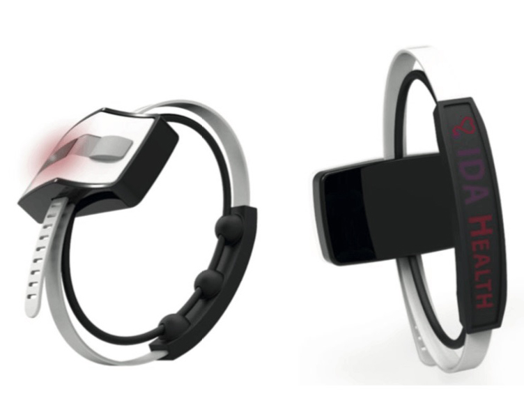
At EuroPCR 2019, Dr Giovanni Amoroso presented a study on an innovative and distinctive medical device, which uses nanotechnology to monitor and manage potential complications in transradial access. The technology is called ROAM (Radial Occlusion Artery Monitoring). It consists of a monitoring bracelet called IdaFlo Tr (IdaHealth, Florida, USA) that allows continuous assessment of arterial flow, with real-time wireless data transmission on arterial compression.
This device can detect abnormal flow in the radial artery after catheterization and warns the doctor or nurse that the radial compression should be adjusted to avoid complete radial artery occlusion. In fact, it is a telemedicine system that transmits collected data on radial artery blood flow to an iPad application. It is a predictive system that helps improve patient safety and reduces the workload on nursing staff.
Key words: transradial access, radial artery occlusion, coronary interventional procedure.

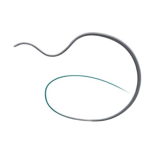
The JUDO guidewires (Boston Scientific) are designed for complex cases of chronic total coronary occlusion and allow maintained direct tactile feedback of the hardness of the plaque. The combination of an effective penetration force, flexibility and precise torque control make them one of the best crossing guidewires particularly for intraluminal crossing of occlusions.
These guidewires are tapered along the last 60 mm, with a core that is surrounded by coiled filaments and goes all the way to the tip. They are hydrophilic-coated to the tip, which measures 0.008”. They possess an innovative technology, called Micro EMT, which means that the core and coil are tapered to match exactly, to enable intraluminal lesion entry. This design avoids exit into the subintimal space; the flexibility of the distal portion along with the precise torque enables effective intraplaque recanalization. These properties reduce whip effect, an important point when aiming for a 3D (3-dimensional) effect, and minimize the possibility of vessel perforation.
Three guidewires of different specifications are available:
- JUDO 1: Soft intraluminal crossing wire for antegrade microchannels, with 1 gf tip load and a penetration force of 31 gf/mm2.
- JUDO 3: Intermediate intraluminal crossing wire for fibrocalcific lesions, 3 gf tip load and a penetration force of 93 gf/mm2.
- JUDO 6: Extra penetration with excellent control in harder lesions, 6 gf tip load and a penetration force of 185 gf/mm2.
Key words: angioplasty guidewire, total chronic occlusion, coronary interventional procedure.
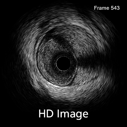
In 2016, 3672 diagnostic intravascular ultrasound (IVUS) studies were performed in Spain. Although the number of cases making use of this technique increased 1.6% compared with 2015, its use has decreased since 2011, when 5124 IVUS studies were performed, in favor of optical coherence tomography (OCT), the use of which has continued to increase.
The quality of intracoronary images acquired with IVUS is improving such that it will undoubtedly affect the choice of diagnostic imaging technique. Several systems provide intracoronary ultrasound images with piezoelectric transducers that range from 20 to 60 MHz. In recent years, there has been an improvement in the axial and lateral spatial resolution of intravascular ultrasound. While axial resolution was previously around 40 microns (even with 60 MHz detectors), with the new OptiCross HD system (Boston Scientific), IVUS resolution approaches that of OCT, which can reach 22 microns. The lateral resolution has also improved, from 80-200 to 50-140 microns, again, approaching that of OCT. This advance is due to a change in the design of the piezoelectric transducer of the catheter, which provides a more sensitive signal with less energy loss and a higher bandwidth, improving the resolution while maintaining the penetration of the IVUS.
This improved image quality makes diagnosis considerably easier and means other diagnostic techniques such as OCT, which require contrast to obtain images, are not required. This is highly relevant in certain patients, such as those with compromised renal function.
It remains to be seen how these changes will affect the rate of use of certain diagnostic imaging techniques over others.
Keywords: ultrasound, IVUS, intravascular ultrasound, HD, OCT, optical coherence tomography

The 2019 Mobile World Congress in Barcelona saw the presentation of an augmented reality system for cardiovascular intervention developed by Philips and Microsoft. The concept of augmented reality is based on the integration of real-time images with live clinical data and 3D medical images to guide procedures accurately and allow the user to concentrate fully on the patient.
This augmented reality system uses images from the new Azurion angiography platform by Philips and a headset with holographic glasses, called HoloLens 2, developed by Microsoft.
Using the HoloLens 2 glasses, Azurion can be controlled in three ways: by voice, eye tracking, or hand gestures. The system allows integration of X-ray and ultrasound images with other 2D and 3D images, meaning that it can be tailored more precisely to the intervention that is to be performed. In reality, HoloLens 2 is a self-contained computer with Wi-Fi connectivity, microphones, spatial audio, electromechanical systems and a power supply.
It is started up with eye movements using Windows Hello and has a holographic visual field that integrates several imaging systems (offline and in real-time) along with 3D clinical data and the controls of the Azurion platform. It safely incorporates Microsoft cloud and artificial intelligence services.
Currently-used systems have large screens showing live 2D images along with other sources of vital data. With this new system it will be possible to work in a 3D augmented reality environment with which doctors will have easy, intuitive and ergonomic control of all available data.
Keywords: Angiography, 3-dimensional imaging, 3D, innovation.