
Innovation in interventional cardiology
High-definition intravascular ultrasound
30/04/2019

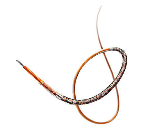
The reSept atrial septal defect occluder (atHeart Medical, Switzerland) is the first occluder to have a bioresorbable structure with no metal components. The frame is made with bioabsorbable filaments that support two polyester patches with radiopaque markers. This frame resorbs over a period of 24 months, while the polyester patches and the radiopaque markers remain at the implantation site.
The device is implanted with a 12-French introducer sheath; currently 3 sizes are available, covering the closure of defects from 4 to 22 mm.
In August 2021, the first 5 patients were treated as part of the ASCENT-ASD clinical trial (NCT04591392) to demonstrate its safety and efficacy in the treatment of ostium secundum-type atrial septal defects compared with the clinical standards of other occluders.
The device is designed as a restorative therapy and is very promising, as it does not leave any metallic structures that could cause future problems.
Keywords: Atrial septal defects, bioresorbable ASD occluder.
With the aim of promoting the fastest and most physiological vascular repair possible, the Evermine stent (Meril Life Sciences, India) has been brought to market, described as an ultrafine stent, measuring 50 micrometers. It is surprising to see how the engineers’ design to obtain the best hemodynamics can be translated to industrial production; I think that we are close to the limits of technological evolution in the field of coronary stents.
The Evermine stent, manufactured from an L605 cobalt-chromium alloy, has a variable strut width as well as a variable crown design to ensure an in vitro radial strength of 1.1 bar. Its coating contains two types of polymers, PLLA and PLGA, which release everolimus at a dose of 1.25 micrograms/mm2. The total thickness of the coating is 2 microns.
This stent can also be considered a hybrid stent, as it has an open-cell design in its middle area, which facilitates access to the lateral branches, and closed cells at the ends to maintain adequate scaffolding and improve its adaptability to the curves of the arteries.
Some very promising preliminary results are already available, from more than 500 patients in 3 studies with follow-up of 1 year: the Evermine 50 EES-1 Study, Evermine 50 EES-KLES Study and Evermine 50 EES-BGM Study.1 It has shown a thrombosis rate of 0% at 6 months and 1 year and a major adverse cardiovascular events (MACE) rate of 2%. These data are consistent with the benefit of a faster endothelialization, with less inflammation and vascular damage, and, from a mechanical point of view, greater ease of implantation, as the device has high flexibility and good transmission of applied force. It comes in a wide selection of sizes, from 2 to 4.5 mm diameter and 8 to 48 mm length.
REFERENCES
1. Sedaghat A, Sinning JM, Werner N et al. In vitro hydrodynamic and acute clinical performance of a novel self-expanding transcatheter heart valve in various surgical bioprostheses. EuroIntervention. 2018;13:2014-2017.
Keywords: Percutaneous coronary intervention, coronary stent.
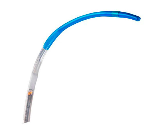
The Dragonfly Opstar catheter (Abbott, USA) is the new generation of catheter for use in coronary imaging with optical coherence tomography (OCT) that brings with it great technological innovation.
The new design of the Dragonfly Opstar catheter combines technological improvements in the tip profile and lens that help easily navigate tortuous anatomy and access distal lesions. This ensures brighter, better quality images to facilitate decision-making.
From a design perspective, the guide wire crossing profile has been reduced by 26% from the previous model and the guide wire port has been reinforced. With these features, the catheter resists 47% more proximal force without torsioning, providing navigability in tortuous anatomies that allows the area of interest to be reached.
There are also technological improvements in the lens, which provides better quality images that are 44% brighter. This makes it easier to detect the external elastic lamina (EEL), enhancing vessel morphology analysis in the vessel to be treated.
Another important point is that the new design of the lens provides better distal visibility, as the lens is positioned 4 mm closer to the tip. This provides an improved focus with the focal point optimized for greater detail in the area of interest.
This new OCT catheter is now available for OPTIS integrated systems, OPTIS mobile systems, ILUMIEN OPTIS systems, and ILUMIEN PCI optimization systems.
Keywords: Optical coherence tomography, coronary artery disease, percutaneous coronary intervention.
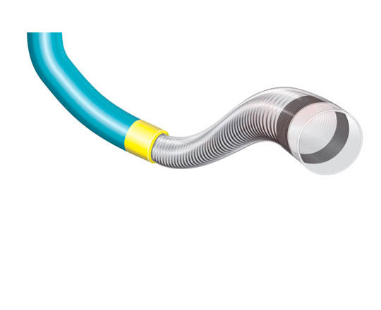
The basic characteristics of a guide catheter extension are navigability, pushability, and the smoothness with which devices can be passed through it.
One of the more interesting innovations in the catalogue of guide catheter extensions is that of the LiquID device (Seigla Medical, USA). Most 6-French extensions have an inner diameter of 0.056’’, while this catheter has an effective inner diameter of 0.061’’. Other available 7-Frech extensions have an inner diameter of 0.062’’ while that of the 7-French LiquID is 0.071’’. These measurements give an idea of the advantages when it comes to working with finer catheters that allow release of large-diameter stents. This can only be achieved with a reduction in the catheter wall thickness, which does not lose its properties thanks to the redesign of the transition zone between the pushing rod and the catheter itself, where the transition is protected by a polymer.
The hydrophilic-coated catheter is 15 cm long, unlike the usual 25 cm, improving navigability in tortuous areas. An interesting feature is that the whole catheter is radiopaque, so the tip does not need any added radiopaque markers.
The catheter is made of a very dense coil that has more volume per millimeter of length, conferring greater resistance to torsion, greater flexibility, and higher radial force. In addition, it has a rounded, atraumatic tip, which guarantees its adaptation to coronary lesions avoids iatrogenic coronary dissection.
Keywords: Guide catheter extension.
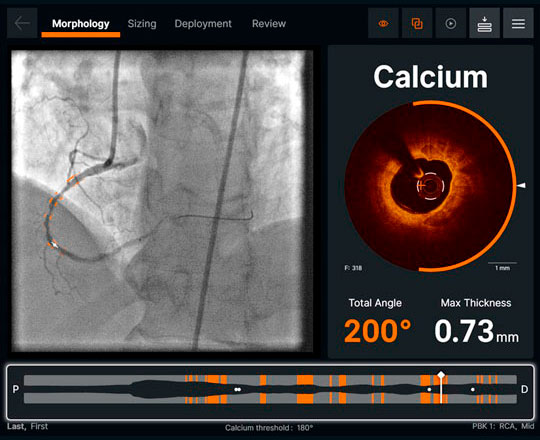
A revolution is coming in the field of image-guided percutaneous coronary intervention, and I think it will mark a new way of working. The use of artificial intelligence models that have been invading all aspects of life in recent years has also reached optical coherence tomography (OCT) consoles via the new software Ultreon 1.0 (Abbott, USA), which has just received the CE mark in Europe in April of this year.
The new Utreon 1.0 software combines OCT with the power of automation driven by artificial intelligence. This improves visualization and allows automation of many measurements that, when done manually, are very time-consuming, to provide precise information and simplify treatment decision-making.
The Ultreon 1.0 software can automatically detect the amount of calcium in the coronary artery and measure the diameter of the vessel to be treated using automatic detection of the external elastic lamina (EEL). This helps improve the accuracy of decision-making during the process of coronary stent implantation in percutaneous coronary intervention.
It also offers an intuitive, simplified workflow that reduces the learning curve and facilitates image interpretation, reducing interobserver variability and increasing diagnostic accuracy and treatment application.
This is the first software of its class to use artificial intelligence and is a great example of technology designed to optimize decision-making, as it reduces uncertainty during the intervention and has a greater accuracy, improving results and patient care.
Keywords: Optical coherence tomography, software, artificial intelligence, percutaneous coronary intervention.