HOW WOULD I APPROACH IT?
The authors present a case of retrogradely uncrossable aortic valve for transcatheter aortic valve implantation (TAVI). This happens with the valve introducer sheath in the femoral artery, and the remaining catheterized accesses. Therefore, a solution to implantation is needed since 1 of the basic steps is missing.
There are 3 situations when crossing a stenosed aortic valve can become especially difficult even for an experienced operator: one is stenosed surgical aortic valves where the ascending aorta is poorly dilated compared to the artificial valve. In this situation, building the latter prevents proper catheter alignment.Another situation is critical aortic stenosis due to small opening orifice. The third situation is bicuspid valves, as it is the case here, with an often dilated ascending aorta or a too vertical valvular plane that complicate maneuvers with the guide catheter. Also, because the bicuspid opening being eccentric often complicates steering the guidewires and the catheters through the valvular orifice.
If we exhaust all retrograde crossing possibilities with different catheters and guidewires, the only option left is antegrade access from the left ventricle (LV) through transseptal catheterization. The use of antegrade access for implantation purposes has already been described in the history of structural heart procedures since it was used for the first TAVI back in 2002.1 Afterwards, it was abandoned due to the high rate of complications and ease of implantation via retrograde transfemoral access. Anyways, some authors still advocate for this access for the lack of better options.2
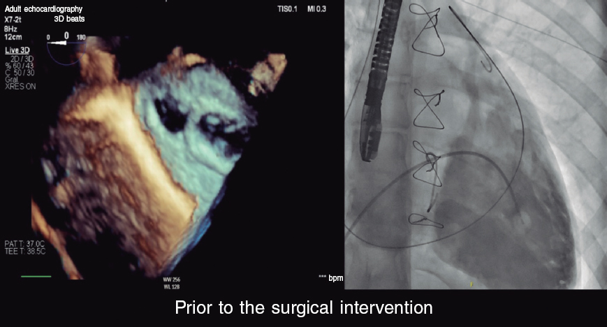
I would perform the procedure using the right femoral vein since it is easier to perform the transseptal access and shorten the procedure since the retrograde access has already been tried for a while; the left femoral vein—already catheterized—is also valid. Currently, transseptal procedures are performed with much safety through transesophageal echocardiography (TEE) guidance. Once the ultrasound-guided right femoral vein has been punctured, a 0.032 in guidewire is advanced across the superior vena cava through which a sheath is advanced for transseptal puncture, often a 63 cm 8-Fr Schwartz SLO (Abbott Vascular, United States). The guidewire is removed and a Brokenburg BRK-1 XS needle is advanced (Abbott Vascular, United States) up to 0.5 cm of the tip of the SLO catheter. At this point, the TEE is performed. At our center—since all procedures are performed under conscious sedation—we would proceed to increase sedation with a bolus of midazolam and use a TEE microprobe that is better tolerated and provides enough imaging for the puncture or else a conventional TEE probe. We will slide from the superior vena cava until the oval fossa and perform the puncture at halfway. Once in the left atrium we direct the transseptal sheath towards the left superior pulmonary vein leaving the 0.032 in guidewire inside. We remove the transseptal sheath and advance a medium curl deflectable Agilis NxT catheter (Abbott Vascular, United States) mounted on it. Once in the left atrium, dilator and guidewire are removed and deflected to bring the catheter closer to the mitral valve. The right anterior oblique view gives us an idea as to where the mitral valve is. Then, the Agilis is turned towards it. A 4-Fr Glidecath multipurpose hydrophilic diagnostic catheter (Terumo Europe, Belgium) is advanced through it until the apex. It bends while being advanced thanks to the Agilis catheter often pointing to the LV outflow tract. A 260 cm J-shaped tip conventional 0.035 in guidewire is advanced until the valve is crossed. Then it’s advanced through the ascending aorta until the abdominal aorta. If crossing is difficult with the multipurpose catheter, a JR4 catheter or a hydrophilic guidewire can be used. From the right femoral artery and through the TAVI introducer, a 6-Fr JR4 catheter we advance a Gooseneck snare of 20 mm-to-25 mm in diameter. The guidewire is captured and then removed through the artery. Therefore, a venoarterial loop has been created. We’ll remove the guidewire as much as possible through the arterial side. From there, we’ll advance the 6-Fr JR4 guide catheter until the LV and loosen up the tension of the loop so that the catheter can be accommodated towards the LV apex. Then, the guidewire is slowly removed from the venous side while keeping the JR4 inside the LV and the high-support guidewire is advanced from the femoral artery. I would keep the Agilis catheter inside the left atrium until to secure the TAVI guidewire into the LV. From that moment onwards, the procedure follows the transfemoral implantation conventional steps.
FUNDING
None whatsoever.
CONFLICTS OF INTEREST
None reported.
REFERENCES
1. Cribier A, Eltchaninoff H, Bash A, et al. Percutaneous transcatheter implantation of an aortic valve prosthesis for calcific aortic stenosis: first human case description. Circulation. 2002;106:3006-3008.
2. Misumida N, Anderson JH, Greason KL, Rihal CS. Antegrade transseptal transcatheter aortic valve replacement: Back to the future? Catheter Cardiovasc Interv. 2020;96:E552-E556.


