
One 72-year-old male who recently suffered from a non–ST-segment elevation myocardial infarction (NSTEMI) underwent a staged percutaneous coronary intervention (PCI) due to significant stenosis of his mid left anterior descending (LAD) and mid left circumflex (LCX) arteries (figure 1A; red arrow). After placing one hydrophilic Hi-Torque intracoronary guidewire into the LCX, one drug-eluting stent (DES) was deployed uneventfully (figure 1B and figure 1C). However, we were unable to extract the guidewire afterwards probably due to entrapment with a calcified plaque. The stronger traction resulted in the partial fracture of the guidewire followed by the disruption of the coils (figure 1D, white arrows; the red arrow points to a second inserted guidewire). Retrieval was unsuccessfully attempted using different techniques like the snare loop technique and the twisting wire technique (video 1 of the supplementary data). After the uncomplicated stenting of the mid LAD, we conducted an optical coherence tomography (OCT). A three-dimensional reconstruction showed remains of the broken wire (figure 2; blue arrows) coming out of the LCX (figure 2; yellow asterisk) and into the left main stem (LMS) and proximal LAD with presence of adhered and free-floating thrombotic material as shown in the cross-sectional views (figure 3A and figure 3B; blue arrows point to the wire remains; the yellow arrow points to the thrombotic material; MLA, minimal lumen area; video 2 of the supplementary data). We immediately proceeded to eliminate the uncoiled filaments from the circulation by deploying one DES into the distal LMS and the proximal LAD. The control OCT conducted showed the wire remains trapped by the struts of the stent against the vessel wall (figure 3C; white arrow: wire remains, highlighted in red-framed box; red asterisks: stent struts).
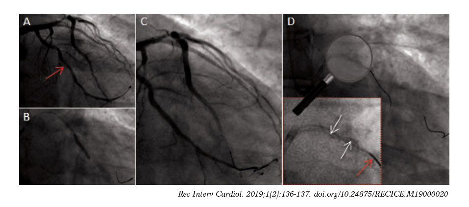
Figure 1
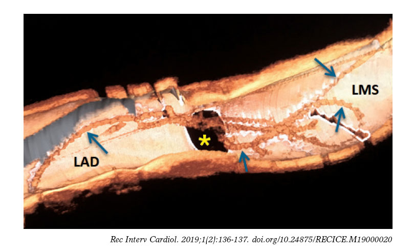
Figure 2
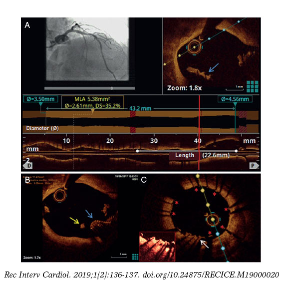
Figure 3
SUPPLEMENTARY DATA
Video 1. Leithold G. DOI: 10.24875/RECICE.M19000020
Video 2. Leithold G. DOI: 10.24875/RECICE.M19000020


