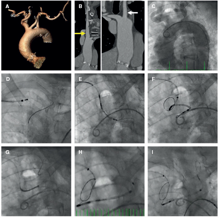
An 84-year-old female with severe aortic stenosis and previous non-disabling stroke was referred to undergo transcatheter aortic valve replacement (TAVR). The 3D computed tomography performed revealed the presence of a type 9 aortic arch with severe tortuosity (figure 1A). It was decided to protect the supra-aortic branches with suitable diameters to be able to use the Sentinel Cerebral Protection System (Boston Scientific, Marlborough, MA, United States). Manipulation length in the left common carotid artery (LCCA) was of, at least, 8 cm which is the distance between the proximal filter and the Sentinel distal edge. Figure 1B: yellow arrow: brachiocephalic trunk, 12 mm-diameter. White arrow: LCCA, 7 mm-diameter. This cerebral protection device (CPD) has a proximal filter for brachiocephalic trunk diameters between 9.0 mm and 15 mm and a distal filter for LCCA diameters between 6.5 mm and 10 mm. The angiography of the aortic arch is shown on figure 1C. This dual-system-filter basket was tried unsuccessfuly over a 0.014 in guidewire despite the use of an articulating sheath (figure 1D-F). After several attempts, a multipurpose catheter was used to engage the LCCA (figure 1G). Using a 300 cm 0.014 in guidewire, the multipurpose catheter was exchanged for the CPD which allowed its suitable deployment (figures 1H,I). The TAVR was performed successfully and the CPD was retrieved (video 1 of the supplementary data). Informed consent was obtained from the patient.
Figure 1.
The major concern is how to balance the risk of stroke after TAVR and the risk of manipulation with guidewires/catheters in supra-aortic arteries. Thus, the rigorous study of the computed tomography scan is the key factor for strategic planning purposes. This was an alternative approach to achieve the placement of a Sentinel device using a multipurpose catheter in a complex aortic arch.
FUNDING
No funding was received for this work.
CONFLICTS OF INTEREST
None declared.
SUPPLEMENTARY DATA
Video 1. Cubero-Gallego H. DOI: 10.24875/RECICE.M20000112


