QUESTION: What are the implications of aortic valve calcification on the outcomes of transcatheter aortic valve implantation (TAVI)?
ANSWER: By design, currently marketed TAVI prostheses require a certain degree of annular calcification to ensure proper fixation. In fact, treating non-calcified valves, such as in pure aortic regurgitation (a scenario for which TAVI has not been approved yet), is associated with a higher risk of malapposition, valve migration, and need for a second prosthesis.1 However, severe valve calcification also poses implantation challenges, as it may compromise the initial procedural success and long-term outcomes.2
Since the planning stage, the presence of severe calcification can hinder the accurate reconstruction of the aortic annular plane and the ability to obtain reliable measurements of its dimensions, thus introducing uncertainty into valve sizing.
Procedurally, severe calcification is associated with a higher risk of immediate complications, such as annular rupture, aortic regurgitation (whether central or paravalvular), and conduction disturbances.3 Furthermore, severe calcification can impede valve crossing and limit the expansion of the prosthesis, which is why valvuloplasty prior to implantation is usually performed to ease valve crossing and allow for greater expansion and better apposition of the prosthesis to the annulus. This increases the number of maneuvers in the aortic root and promotes the embolization of debris, which is associated with a higher risk of ischemic events.
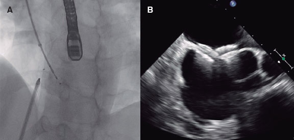
Severe calcification can limit the expansion of the prosthesis and alter leaflet configuration (the so-called pin-wheeling phenomenon), which increases leaflet stress and has been associated with in vitro studies with reduced durability. Furthermore, underexpansion is associated with elevated gradients and a higher rate of central (due to leaflet distortion) and paravalvular (due to annular malapposition) regurgitation.
Q.: What morphological and quantitative aspects of valvular calcification do you assess at your center?
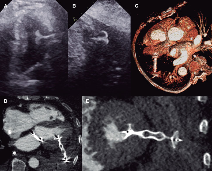
A.: The gold standard for quantifying valvular calcium is computed tomography (CT), which uses either the Agatston score from non-contrast scans or calcium volume from contrast-enhanced CT angiography.4 The degree of calcification, its location, morphology, and distribution asymmetry are key determinants of procedural success and risk of complications. Several studies have associated moderate or severe calcification at the landing zone with a higher risk of paravalvular regurgitation, conduction disturbances, and annular rupture.3 It seems that calcifications protruding into the lumen are most associated with complications, while flatter calcifications have less impact. In addition, severe commissural calcification has been associated with residual regurgitation in that region too.5
The predominant location of calcification on the leaflets body is related to the degree of underexpansion and the prosthesis functionality, which can also influence the risk of coronary compromise in cases where leaflets are calcified at the level of the coronary ostium, especially with low-lying coronary arteries and narrow sinuses.
Ultimately, the interaction between calcium and prosthesis depends on the type of device. Thus, supra-annular prostheses better preserve the geometry of the leaflet when calcification is located at the annulus or left ventricular outflow tract (LVOT), while prostheses with narrower waists and lower radial forces result in less displacement of the leaflets toward the coronaries.
Calcium eccentricity is a major challenge, and the morphology of the valve plays a key role here: bicuspid valves usually show more complex calcification patterns than tricuspid valves do, with greater asymmetry and often calcified raphes that promote asymmetric expansion and prosthesis displacement toward areas of lower resistance, which are features that can also hinder positioning and stable release.
In summary, beyond describing the degree of calcification, what we need to do is target its location (annulus, LVOT, leaflet body, commissures, extension to the sinotubular junction, prominent nodules facing the coronary origins, etc.), the presence of nodules protruding into the lumen or flatter annular-aligned calcifications, as well as their eccentricity.
Q.: How does the degree of calcification affect valve type selection?
A.: Severe calcification increases the risk of complications, particularly paravalvular regurgitation, annular rupture, underexpansion, and conduction disturbances.3 We, therefore, try to select the prosthesis that best matches each case, based on calcium location and characteristics.
Generally, we prefer self-expanding valves and avoid aggressive pre- and post-dilatations in cases with prominent calcium nodules, as their interaction with the balloon increases the risk of dissection or rupture, which are complications with an associated mortality rate close to 100%. Another less common scenario where balloon- expandable valves should be avoided is a small, calcified sinotubular junction relative to the annular size, due to the risk of balloon- induced injury or aortic dissection.
Self-expanding valves, with a more gradual release than balloon- expandable ones, theoretically offer better adaptability to irregular anatomies and may be preferable in annuli with very irregular calcification. However, this is a complex trade-off, as they provide less complete sealing due to their lower radial force but still may reduce the risk of rupture. Balloon-expandable valves may be a very good option when calcification is limited to a specific annular area, especially if it does not protrude excessively.
Additionally, in the presence of significant calcification, it is essential to favor prostheses with an outer sealing skirt and high radial force.
Since LVOT calcification increases the risk of conduction disturbances, we often opt for recapturable valves to optimize final positioning.
In conclusion, valve choice seeks to balance the risks of regurgitation, conduction disturbances, aortic complications, and residual gradients.6
Q.: And how does the degree of calcification affect valve sizing?
A.: Calcification of the leaflet base and LVOT complicates the identification of the cusp nadirs and annular reconstruction, impeding accurate annular boundary definition and precise measurement. Since valve sizing relies primarily on these measurements, excessive calcification may lead to under- or oversizing.
Excessive calcification clearly impacts balloon-expandable valve sizing, generally leading to smaller oversizing (by reducing balloon inflation volume or valve size). However, its influence is not as direct in self-expanding valves where the risk of aortic rupture or dissection is lower due to their reduced radial force, meaning we can aim for standard oversizing or, in cases of borderline annuli between 2 valve sizes, even slightly larger. This is done to achieve better sealing, provided that the anatomy of the sinuses of Valsalva allows for adequate leaflet expansion without a higher risk of coronary compromise. Furthermore, while slight undersizing of balloon-expandable valves can be mitigated by adding a few milliliters of volume to the balloon without compromising its functionality, the problem of an undersized self-expanding valve has no solution because the nitinol material of the valves recovers its factory design at body temperature, even after aggressive post-dilation, which, in turn, perpetuates the problem.
On the other hand, calcium may prevent the adequate expansion and apposition of an oversized valve, thus degrading procedural outcomes and making sizing decisions particularly difficult in many cases.
Q.: Does the procedure differ based on valve calcification?
A.: In cases of severe calcification, we always perform predilatation regardless of the type of valve we’ll be using. Postdilatation is also frequently required, and often more aggressive, to optimize the degree of regurgitation.7
Although optimal positioning is always desired, it is especially critical in “champagne cork”–shaped prostheses designed to achieve optimal oversizing at a certain implant depth; deeper positioning reduces oversizing and increases the risk of regurgitation.
In cases of LVOT calcification, repeated valve movement in and out of the ventricle should be avoided to reduce the risk of conduction system injury.
Finally, significant underexpansion may lead to hemodynamic instability until adequate expansion is achieved via postdilatation. Therefore, one must be ready to support the patient hemodynamically while performing these maneuvers. In this situation, it is of paramount importance to maintain ventricular guidewire access, as crossing a severely underexpanded valve may be difficult. In cases of severe hemodynamic compromise before release, it may be advisable to recapture (if the system allows it) the valve and perform a more aggressive valvuloplasty.
Q.: What are the advantages of self-expanding prostheses in the most severely calcified valves?
A.: In these prostheses, expansion occurs gradually, allowing progressive adaptation to annular irregularities. However, a key limitation compared with balloon-expandable valves is their lower radial force, which increases the risk of regurgitation (a problem noted since the early days of the technique). Next-generation prostheses include features to mitigate this risk. Many incorporate an outer skirt to improve sealing and reduce paravalvular regurgitation. Moreover, several self-expanding valves are now recapturable, allowing assessment of implant height and regurgitation before full release, and repositioning if necessary. Release systems now offer greater stability, more predictable deployment, easier positioning, and fewer recaptures and manipulations in the aortic root.
Secondly, in supra-annular self-expanding valves, normal leaflet configuration is unaffected by calcification at the annulus or LVOT, given their higher positioning, thus generally preserving valve function and excellent hemodynamics even with significant calcification.
Moreover, in severe calcification, the risk of aortic dissection or annular rupture (complications whose mortality rate is close to 100%) is primarily linked to balloon expansion. In this context, a self-expanding valve protects against such complications, provided aggressive pre- or post-dilatation is avoided.
Lastly, self-expanding prostheses typically have a better profile than balloon-expandable ones, enhancing trackability and crossing ability, and facilitating the procedure.
Q.: Are all prostheses the same in this context?
A.: First-generation self-expanding valves were associated with higher rates of paravalvular regurgitation, malapposition, embolization, and need for a second valve in moderate-to-severe calcification. These outcomes have improved parallel to the technical advancements made in the latest versions,8 such as repositionability, recapture capability, and outer skirts. Therefore, in severe calcification, if a self-expanding valve is going to be used, one should prefer models with higher radial force, outer skirt, and repositioning and recapture capabilities. It is essential to remember that even within the same type of valve, several models can have different characteristics that must be well understood.
FUNDING
None declared.
STATEMENT ON THE USE OF ARTIFICIAL INTELLIGENCE
Artificial intelligence was not used.
CONFLICTS OF INTEREST
R. del Valle reports having received speaker fees from Medtronic Ibérica and Mercé V for educational courses, and from Medtronic International for consultancy work.
REFERENCES
1. Yoon SH, Schmidt T, Bleiziffer S, et al. Transcatheter Aortic Valve Replacement in Pure Native Aortic Valve Regurgitation. J Am Coll Cardiol. 2017;70:2752-2763.
2. Alperi A, Del Valle R, Avanzas P. Impact of aortic valve calcification on TAVI. Should we rethink existing concepts?Rev Esp Cardiol. 2025;78:519-520.
3. Okuno T, Asami M, Heg D, et al. Impact of Left Ventricular Outflow Tract Calcification on Procedural Outcomes After Transcatheter Aortic Valve Replacement. JACC Cardiovasc Interv. 2020;10:1789-1799.
4. Flores-Umanzor E, Keshvara R, Reza S, et al. A systematic review of contrast-enhanced computed tomography calcium scoring methodologies and impact of aortic valve calcium burden on TAVI clinical outcomes. J Cardiovasc Comput Tomogr. 2023;17:373-383.
5. Ewe SH, Ng AC, Schuijf JD, et al. Location and severity of aortic valve calcium and implications for aortic regurgitation after transcatheter aortic valve implantation. Am J Cardiol. 2011;108:1470-1477.
6. Kim WK, Blumenstein J, Liebetrau C, et al. Comparison of outcomes using balloon-expandable versus self-expanding transcatheter prostheses according to the extent of aortic valve calcification. Clin Res Cardiol. 2017;106:995-1004.
7. John D, Buellesfeld L, Yuecel S, et al. Correlation of device landing zone calcification and acute procedural success in patients undergoing transcatheter aortic valve implantations with the self-expanding CoreValve prosthesis. JACC Cardiovasc Interv. 2010;3:233-243.
8. Farhan S, Stachel G, Desch S, et al. Impact of moderate or severe left ventricular outflow tract calcification on clinical outcomes of patients with severe aortic stenosis undergoing transcatheter aortic valve implantation with self- and balloon-expandable valves:a post hoc analysis from the SOLVE-TAVI trial. EuroIntervention. 2022;18:759-768.
* Corresponding author.
E-mail address: raqueldelvalle@gmail.com (R. del Valle Fernández).


