HOW WOULD I APPROACH IT?
Authors present an interesting case of a 10-year-old with a familial history of Rendu-Osler-Weber disease. The patient shows signs of cyanosis, polyglobulia, and baseline oxygen saturation levels of 85% due to a large and complex pulmonary arteriovenous malformation (PAVM) that is causing systemic desaturation due to the existence of a significant right-to-left shunt.
PAVMs are direct connections between arterial branches—often the pulmonary artery—and pulmonary veins without a normal capillary bed connected through an aneurysmal sac that can be partially septated in the inside. PAVMs can be categorized into simple, when they receive blood from a single afferent arterial vessel, and complex, when afference is multiple.
Most of them are congenital and in over 70% of the cases they are associated with Rendu-Osler-Weber disease or hereditary hemorrhagic telangiectasia. Although it is a dominant autosomal disease that can be diagnosed through a genetic study the so-called Curaçao diagnostic criteria1 are often used to achieve diagnosis:
Recurrent epistaxis.
Multiple telangiectasias in typical locations: lips, oral cavity, fingers, and nose.
Visceral vascular malformations: gastrointestinal, pulmonary, hepatic, cerebral or spinal.
First-degree relative who is a disease carrier.
With 3 or more criteria, the diagnosis becomes conclusive. With just 2 the diagnosis is possible.
Most of the times this disease is asymptomatic, and the presence of clinical signs depends on the size and number of these signs. The symptoms surrounding PAVMs are:
Associated with desaturation—due to the right-to-left shunt—with peripheral oxygen saturation levels < 90% that lead to cyanosis, acropachies, and reactive polycythemia.
Due to the frailty of the PAVM walls that can rupture towards the bronchial bed causing hemoptysis or towards the pleural space causing hemothorax.
Due to the lack of pulmonary capillary filtration, paradoxical embolisms can occur and, sometimes, be followed by the formation of cerebral abscesses, which is not rare.
Diagnosis is based on the existence of suggestive clinical signs. Thoracic x-rays show disturbances in over 95% of the patients. The agitated saline contrast echocardiography reveals the presence of bubbles passing through after 3-5 seconds (3 to 8 cardiac cycles). Currently, the computed tomography scan is the reference imaging modality; it allows accurate anatomical studies, and is used to plan embolization, as well as for evolutionary follow-up purposes.
Although, initially, surgical resection was indicated, the treatment of choice is endovascular embolization. It is indicated2 when the afferent arterial vessel is ≥ 3 mm because with this size the risk of paradoxical embolism is higher compared to the annual 1.5% reported. Thanks to state-of-the-art materials the procedure can be performed safely in afferent arterial vessels < 2 mm, which is why each patient should be assessed individually when the size of the arterial vessels is between 2 mm and 3 mm.
Embolization can be performed using metallic coils as described by Gianturco et al.3 in 1975. In our case we used the MReye Flipper Detachable Embolization Coil and Delivery System (Cook, United States). To start embolization with the right controlled delivery coil, the size of the coil should be, at least, 30% larger compared to the target vessel diameter. Also, it should be released into the afferent arterial vessel as distal as possible. Other coils are deployed following the first coil until the occlusion is complete. Another option is to use nitinol vascular plugs for embolization purposes. In our case we used devices from the Amplatzer Family of Vascular Plugs (Abbott, United States). The size selected was 30% to 50% larger compared to the diameter of the target vessel, and the plug was deployed into the afferent arterial vessel as distal as possible to not interfere with other branches leading towards the healthy parenchyma. Choosing one material over the other often depends on the operator’s experience and preferences.
In the case presented here, the indication for closure is a consequence of the significance of the shunt that is causing desaturation. It is also associated with the complications occurred due to the size of the malformation, the paradoxical embolism, and eventual rupture.
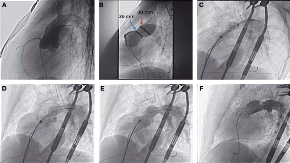
In our own experience, analyzing the computed tomography scan beforehand allows us to study embolization-eligible regions, choose the devices that will be used, and the most favorable angles to work with. It also allows us to keep an eye on the presence of branches perfusing healthy tissue so they don’t interfere with the devices.
The procedure should be performed under general anesthesia via right femoral venous access using a 6-Fr introducer sheath to advance a Wedge catheter (Teleflex, United States) towards the right pulmonary branch. From that position, it should be advanced selectively towards the afferent arterial vessels while supported by 4- or 5-Fr Judkins Right 4.0, Cobra or Vertebral multipurpose diagnostic catheters mounted over 0.018 to 0.035 in hydrophilic guidewires (Radiofocus Guide Wire, Terumo, Japan) or 0.014 in workhorse coronary guidewires. Selective angiographies are performed in each afferent arterial vessel to confirm the characteristics of the embolization target vessel. Once it reaches the niche of the malformation, the transporter sheath is exchanged. In this context, my preference is to use nitinol self-expanding devices like the Amplatzer Vascular Plug (AVP), the Amplatzer Vascular Plug II (AVP II) or the Amplatzer Vascular Plug IV (AVP 4) (Abbott, United States). There are times that we use coils and plugs in the same patient based on the anatomical characteristics of each afferent arterial vessel. In this case I would approach the 3 afferent arterial vessels one by one within the same procedure. Then, I would deploy the devices after confirming their correct position, and stability. Given the lack of a limiting capillary bed, device migration to the left cavities can occur. Therefore, all devices should be properly sized. If this complication occurs the device can be reached via arterial access, often femoral, captured with a snare, and then removed with a larger 2-Fr sheath compared to the one used for release.
After the occlusion—during the evolutionary follow-up—it is advisable to perform a computed tomography scan after 6 months to confirm the effective closure and discard the recanalization of embolized arteries or the appearance of new malformations. Similarly, periodic controls are advised.
FUNDING
None.
CONFLICTS OF INTEREST
None whatsoever.
REFERENCES
1. Shovlin CL, Guttmacher AE, Buscarini E, et al. Diagnostic criteria for hereditary hemorrhagic telangiectasia (Rendu-Osler-Weber syndrome). Am J Med Genet. 2000;91:66-67.
2. Faughnan ME, Mager JJ, Hetts SW, et al. Second International Guidelines for the Diagnosis and Management of Hereditary Hemorrhagic Telangiectasia. Ann Intern Med. 2020;173:989-1001.
3. Gianturco C, Anderson JH, Wallace S. Mechanical devices for arterial occlusion. Am J Roentgenol Radium Ther Nucl aMed. 1975;124:428-435.
* Corresponding author: Servicio de Cardiología, Hospital Universitario Cruces, Pza. de Cruces s/n, 48903 Barakaldo, Bizkaia, Spain.
E-mail address: blancomata@yahoo.es (R. Blanco Mata).


