Mitral regurgitation (MR) is one of the most prevalent valvular heart diseases in the world.1 Although surgical mitral valve replacement has shown to improve clinical outcomes in patients with severe primary MR, surgery is still being denied in a significant number of patients due to their multiple clinical comorbidities.2 The current clinical practice guidelines for the management of severe secondary MR recommend the surgical valve intervention only in cases where concomitant coronary revascularization is indicated.3
Transcatheter mitral valve repair using the edge-to-edge technique (MitraClip, Abbott Menlo Park, CA, United States) has shown to improve quality of life and reduce all-cause mortality in patients with heart failure and secondary MR refractory to the optimal medical treatment.4 However, the level of MR reduction achieved by MitraClip is inferior compared to the surgical techniques, and its overall use is limited by several anatomical factors.5 Transcatheter mitral valve implantation is emerging as a potential therapeutic alternative that could overcome some of the current limitations of edge-to-edge repair.6 Due to their technical design features, most transcatheter mitral valve implantation systems use the surgical transapical approach. Limited experience has been gathered on the use of dedicated transseptal systems. Finally, complex anatomical features such as the possibility of left ventricular outflow tract obstruction and presence of mitral annular calcification have limited the fast clinical adoption of this technology.
The field of catheter-based mitral valve intervention is rapidly expanding, and translational experimental models are seriously needed for the proper validation of these technologies. Unlike aortic valve disease, MR is due to several pathological conditions that result in different anatomical substrates that are not easy to reproduce in experimental models. Catheter-based technologies are designed taking into consideration specific anatomical targets such as annular dilatation or chord elongation, which are also challenging to reproduce in experimental animal models. Significant differences exist between humans and animal models. In the first place, one of the most important challenges we face is annular size. Devices developed for human use are typically larger compared to the annulus seen in common experimental models, which at times, requires developing customized valve sizes. Secondly, the aortomitral curtain is particularly small, which often leads to device interaction with the aortic valve. Thirdly, the mitral tissue is thin and friable providing little support to technologies that require the use of anchors or pads to remain in position. Finally, the left atrium is flat and shallow and provides little room for the validation of technologies via transseptal access.
The use of diseased animal models is not typically required to validate structural heart technologies and most of the validation work can be done on the bench or on healthy animal models. Healing and thrombogenicity of valve materials is particularly important and can be validated in healthy animals. The stability of the frame and durability of the leaflets can also be tested and is of particular significance in the transcatheter mitral valve implantation space. The mechanism of the deployment and delivery system can also be tested but, overall, the retention of the valve depends on the mechanism of anchoring used, which could be challenging due to the lack of structural support. In these cases, the surgical placement of the valves is needed for the long-term stability of the implant.
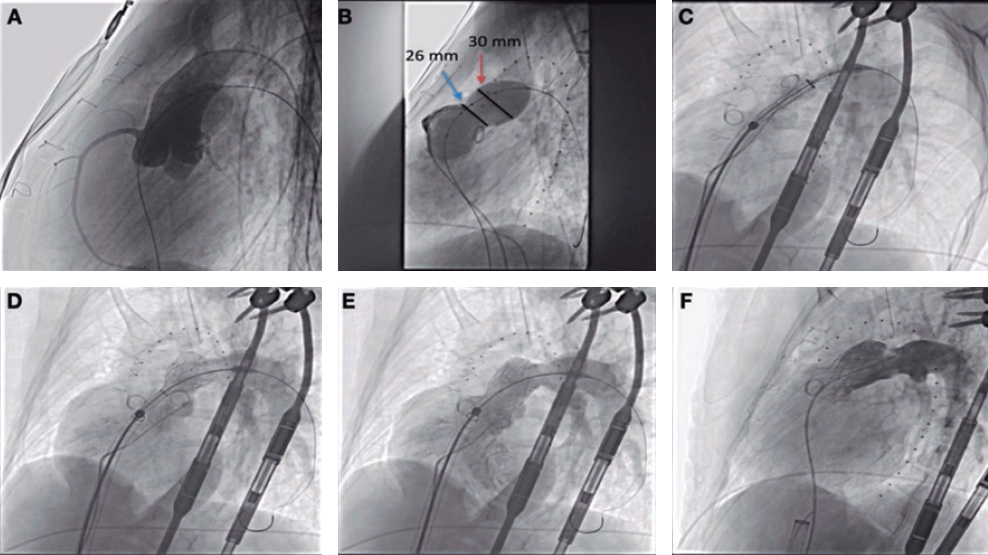
The use of diseased animal models is often spared to assess the efficacy of the device or test particular device features (ie, anchoring). Several animal species, primarily dogs suffer from primary MR due to leaflet prolapse and have been used to test several catheter-based technologies.7 However, these models are expensive and difficult to provide. Several groups have already developed secondary MR models by inducing ischemia of the posteromedial papillary muscle.8-9 These models have resulted in a high periprocedural mortality rate and moderate levels of clinically relevant MR. The study conducted by Rodríguez-Santamarta et al. recently published on REC: Interventional Cardiology presents a variation of this model by adding volume overload and creating an aorto-pulmonary fistula following the myocardial infarction of the circumflex artery.10 The number of animals was small, but researchers were able to prove the feasibility of model development. In this study, the level of MR was moderate (at most) and other features associated with MR such as annular dilatation were present. These morphological features, although obvious on the image assessment, were subtle in nature and probably in their early stages compared to patients who suffer from severe MR.
The field of structural heart procedures is changing very rapidly, and experimental models are essential for the proper validation of these technologies. Healthy animal models are perhaps enough to test the mechanism of the device delivery system, healing, and durability. Diseased animal models can help validate device efficacy, mechanisms of anchoring, and the long-term stability of the device. However, due to the high anatomical variability seen in humans compared to animals, long-term results may be confusing and require careful analysis by multidisciplinary teams before starting the first tests in humans. A multi-modality approach is highly desirable in the validation process of structural heart technologies. Although animal data are key, proper validation including human tissue and imaging correlation studies may help minimize the misinterpretation of experimental signals and define the developmental pathway of structural heart technologies.
FUNDING
No funding was received for this work.
CONFLICTS OF INTEREST
J. Granada is co-founder of Cephea Valve Technologies.
REFERENCES
1. Nkomo VT, Gardin JM, Skelton TN, Gottdiener JS, Scott CG, Enriquez-Sarano M. Burden of valvular heart diseases:a population-based study. Lancet. 2006;368:1005-1011.
2. Mirabel M, Iung B, Baron G, et al. What are the characteristics of patients with severe, symptomatic, mitral regurgitation who are denied surgery?Eur Heart J. 2007;28:1358-1365.
3. Nishimura RA, Otto CM, Bonow RO, Carabello BA, Erwin JP, Fleisher LA, et al. 2017 AHA/ACC Focused Update of the 2014 AHA/ACC Guideline for the Management of Patients with Valvular Heart Disease:A Report of the American College of Cardiology/American Heart Association Task Force on Clinical Practice Guidelines. Circulation. 2017;135:1159-1195.
4. Stone GW, Lindenfeld JA, Abraham WT, et al. Transcatheter mitral-valve repair in patients with heart failure. N Engl J Med. 2018;379:2307-2318.
5. Boekstegers P, Hausleiter J, Baldus S, et al. Percutaneous interventional mitral regurgitation treatment using the Mitra-Clip system. Clin Res Cardiol. 2014;103:85-96.
6. Del Val D, Ferreira-Neto AN, Wintzer-Wehekind J, et al. Early Experience With Transcatheter Mitral Valve Replacement:A Systematic Review. J Am Heart Assoc. 2019;8:e013332.
7. Morgan KRS, Monteith G, Raheb S, Colpitts M, Fonfara S. Echocardiographic parameters for the assessment of congestive heart failure in dogs with myxomatous mitral valve disease and moderate to severe mitral regurgitation. Vet J. 2020;263:105518.
8. Pasrija C, Quinn RW, Alkhatib H, et al. Development of a Reproducible Swine Model of Chronic Ischemic Mitral Regurgitation:Lessons Learned. Ann Thorac Surg. 2020;111:117-125.
9. Hamza O, Kiss A, Kramer AM, Tillmann KE, Podesser BK. A novel percutaneous closed chest swine model of ischaemic mitral regurgitation guided by contrast echocardiography. EuroIntervention. 2020;16:e518-e522.
10. Rodríguez-Santamarta M, Estévez-Loureiro R, Pérez Martínez C, et al. Experimental model of mitral regurgitation in a porcine model. REC Interv Cardiol. 2021;1:12-18.
Corresponding author: CRF-Skirball Center for Innovation, Cardiovascular Research Foundation, 8 Corporate Dr, Orangeburg, New York 10962, United States.a
E-mail address: jgranada@crf.org.


